Radiologist Interview Series – Prof. Hildo J. Lamb
2023-02-13

The radiologist as a healthcare navigator

Interview with Prof. Hildo J. Lamb, M.D., Professor of Radiology and Director of Cardiovascular Imaging at Leiden University Medical Center.

Professor Lamb, how long have you been working with SEARCH Lung CT?

Prof. Dr. Hildo J. Lamb: We first started talking to contextflow about three years ago. This then turned into a partnership. We have been working together intensively for about two years and continue to develop the solutions together.

How should I understand this cooperation?

Prof. H. J. Lamb: One of the challenges in radiology, for example, is the integration of AI solutions into reporting systems. We work with a system from Sectra that is open to the integration of third-party systems. We have thus managed to integrate contextflow’s AI solution into the workflow and are now using it in routine clinical practice.

We are currently working with SEARCH Lung CT and want to use the software to identify abnormal findings in CT images. We expect this to lead to better workflow management. In the morning, we start well-rested and fresh with abnormal, complex cases. In the afternoon, when a certain fatigue sets in, the cases classified as normal by the artificial intelligence (AI) are then assessed. We hope this approach will result in safer and better reporting.

Are there already further plans with SEARCH Lung CT?

Prof. H. J. Lamb: Oh yes. When all radiologists are convinced that this AI solution will support them effectively, we will use it in the long-term, for example, to calculate lung nodule volume or volume doubling times. That will be another leap in quality. Currently, due to time constraints, we only look at the last preliminary scan. It’s impossible to manually quantify all the scans over the course of a lung cancer treatment. AI can do that. With all the automated tools, we can create a curve from each data point and track the change in nodule volume. Today, we only measure the diameter of the lesions. But it is better to look at the volume because that is much more sensitive to changes over time. That should be the next step with SEARCH Lung CT.

You talk a lot about therapy management and your roles in that. Where do you see the role of the radiologist in the future?

Prof. H. J. Lamb: We are faced with the challenge that both the image data volume of the images and the complexity of the cases are constantly increasing. At the same time, the need for quantification – here in particular of volumes, less so of diameters – is increasing because we cannot evaluate lesions visually alone. All in all, this leads to an exponential increase in our workload. The dangers range from incorrect reporting to burnout. I am convinced that AI can help us immensely. It is predestined to detect lesions and quantify them automatically. Importantly, we still need a radiologist to interpret this quantification. This is exactly where I see the future of our discipline: in the interpretation of findings, in explaining the results against the background of the patient’s entire medical history, and in advising on the next steps.

In other words, the radiologist as a pilot.

Prof. H. J. Lamb: We actually see the radiologist as a navigator of health care. We want to be the spider in the web of healthcare, with an integrated view of the patient. So we’re not just looking at the heart or just the lungs or just the brain, but the whole body. We need to leave behind specialization and niche thinking and look at the patient as a whole. To do this, however, we need time, and AI-supported systems can give us that by taking over time-consuming routine activities.

How far away is this scenario?

Prof. H. J. Lamb: It’s hard to say. First of all, we have to develop all the necessary technologies. Then they have to be reliable and validated so that they work smoothly. That will certainly take a few more years. I think that in five years we will have some kind of basic arsenal of techniques to do this. And then the transition has to start, that we embrace it and accept the integration of AI.

What impact will AI have on the work of the radiologist then?

Prof. H. J. Lamb: The workflow for radiologists is going to change over the next five to 10 years. That is why it is so important that we get involved now, at the beginning of this change. We can’t just wait and see what the engineers and companies do. Rather, we need to take the lead in this change. We need to define what our future looks like.

That brings us back to the topic of collaboration with companies. What should this look like?

Prof. H. J. Lamb: The best model we have experienced is to develop a product together in co-creation. If a solution is designed in an ivory tower, it will never meet our requirements. The key to successful and accepted AI solutions therefore lies in close cooperation between users and companies.

Thank you very much for the exciting and inspiring insights, Professor Lamb.

Other News

Radiologist Interview Series – Prof. Dr. Elmar Kotter & PD Dr. Daniel Pinto dos Santos
2023-02-07

February is known as the month of love, so we are sharing our affection for radiologist friends with an interview series. Each Tuesday this month, we’ll release another talk with a distinguished advisor or expert on various topics surrounding AI in radiology.

Up first is actually a dynamic duo: Prof. Dr. Elmar Kotter, Senior Physician, IT & QM in the Department of Radiology at the University Hospital Freiburg, and PD Dr. Daniel Pinto dos Santos, Senior Physician at the Institute for Diagnostic and Interventional Radiology at the University Hospital Cologne and at the Institute for Diagnostic and Interventional Radiology at the University Hospital Frankfurt.

To begin with, a basic question: What is your opinion of artificial intelligence (AI) in radiology?

Dr. Daniel Pinto dos Santos: For me, it is impossible to imagine radiology without AI. There will be many use cases in the future where it will become very relevant. AI is here to stay – and rightly so.

What would be predestined use cases?

Prof. Dr. Elmar Kotter: You can distinguish between low complexity, high volume use cases and high complexity, low volume use cases. A typical use case that comes to mind spontaneously is fracture detection. Or in the emergency room as a safety check that no pulmonary artery emboli are missed. Here, AI can be of proven help.

So it’s primarily about safety, about a “second opinion.” Or do you see AI also in primary diagnosis?

Prof. E. Kotter: I see the latter rather critically. The way we are set up today, the radiologist has to decide in the end. There can certainly be a preliminary diagnosis by AI, which presents its results. But AI is still a long way from being able to make decisions on its own.

Do we want to get there?

Dr. D. Pinto dos Santos: That opens up a very difficult ethical and legal discussion, which I also don’t see as a priority at all right now. I see AI solutions as absolutely sensible for safeguarding diagnostics. But I also see another aspect: There are many things that we radiologists don’t do today because they are too time-consuming in everyday clinical practice, for example, recording organ sizes or lung changes. I see the greatest potential for AI in these use cases, namely to make information available to us that we would always have liked to have, but which we do not currently have.

Prof. E. Kotter: I fully agree with Daniel. Collecting quantitative data automatically and systematically is an important use case for AI. Aortic diameters, for example, we only measure today if they seem conspicuous or because we are asked to do so. But we don’t do that systematically. Measuring bone density or measuring liver density are all applications that would certainly be very helpful.

In order to be able to use the results easily, they have to be integrated into the reporting systems, i.e., into the PACS. Is that also your main requirement?

Prof. E. Kotter: Of course, but that is already reality. Ready-prepared images are sent to the PACS, and the radiologist can look at them there. This data exchange between different applications is standardized via corresponding IHE protocols.

Dr. D. Pinto dos Santos: I am convinced that the integration of the systems will make the difference between an accepted and an unaccepted solution in the future.

Prof. E. Kotter: The ideal is that we will eventually work with report templates and that the AI algorithms will help us fill in these templates. The radiologist fills in the gaps. I can well imagine that different algorithms will work together to detect different things and that, in the end, all the information will be summarized in a more structured report.

What does integration look like with contextflow?

Prof. E. Kotter: In addition to the variant just described, there is also the option of working directly in the contextflow interface. This is a bit more demanding but makes sense, for example, for decision support – especially with the connection to STATdx. A third scenario is the quality case application: Here, the software compares the results it has obtained from the images with the radiologist’s findings and looks to see if there are any mismatches. If there are, the software sends an email to the radiologist with a corresponding note.

You both worked on the new version of contextflow’s software. What was that process like?

Dr. D. Pinto dos Santos: I received screenshots or mock-ups with very specific questions: How do you see this feature? What representation would you like to see so that the software is intuitive to understand and use? That was a fruitful exchange that I really enjoyed. I think this involvement in development processes is enormously important because you have to understand each other in order to create a good tool that is helpful in everyday life.

Prof. E. Kotter: I would also like to praise the contextflow team. I experienced the exchange as a good, constructive discussion. Especially my feedback, when I tested the system, I perceived it as very welcome. It is not the case with all companies that criticism really falls on fertile ground. But this is the only way to move a solution forward.

What is your experience with contextflow solutions?

Dr. D. Pinto dos Santos: I think they can do some things very well, for example quantifying change, that’s a real plus. Personally, I think it’s very efficient in that it’s less about prescribing a diagnosis and more about helping to find image patterns. That enables the radiologist to make a better diagnosis.

Prof. E. Kotter: I’d like to pick up on that because I really see contextflow as unique in that point. I don’t know of any other solution that offers me comparable cases and thus supports me in my decision making. The solution also very elegantly circumvents the frequently cited problem of AI as a black box, i.e. the lack of transparency in decision making.

Professor Kotter, Doctor Pinto dos Santos, thank you very much for taking us a little way into the co-development process of contextflow.

Other News

Collective Minds Radiology & contextflow partner to enable testing of AI without barriers
2023-02-01

Immediate access via Collective Minds Radiology’s portal enables radiologists to upload cases and quickly receive contextflow results

Vienna, 10, January, 2023: Healthcare collaborators Collective Minds Radiology and chest CT experts contextflow GmbH have partnered to help radiologists evaluating complex suspected ILD, COPD, and lung cancer cases. Specifically, radiologists can securely upload chest CTs to the free Collective Minds platform, and contextflow will analyze the images and provide them with quantitative results without the need for a lengthy software integration process. 

Based in Stockholm, Sweden, Collective Minds Radiology offers the world’s largest cloud-based collaboration platform for radiologists. This global community service enables radiologists to share interesting cases, along with their clinical questions, and then to receive insights back from other practicing radiologists. This collective intelligence can be particularly helpful when a radiologist is uncertain of an imaging findings. The end goal being, of course, to help patients get their correct diagnosis faster.

contextflow offers comprehensive computer-aided detection software for chest CT to support the diagnosis of suspected ILD, COPD and lung cancer. The company’s core technology, ADVANCE Chest CT, automatically detects, quantifies and visualizes 8 disease patterns and lung nodules in CTs of the lungs, displaying relevant information directly in the radiologist’s PACS viewer. In addition, contextflow’s TIMELINE feature quickly and objectively tracks changes in lung nodules over time, currently impossible in the given radiology workflow.

Regarding the announcement, Collective Minds Radiology CEO & Co-Founder Anders Nordell says “We are thrilled to announce our partnership with contextflow, a leading provider of AI-powered diagnostic tools for radiology. This partnership allows us to enrich our healthcare collaboration services with cutting-edge diagnostic capabilities, further enhancing our ability to support our community of healthcare professionals around the world. We believe that this partnership will have a positive impact on our customers and ultimately improve patient outcomes.”

contextflow Chief Commercial Officer Marcel Wassink continues, “Implementation of AI tools has so far been a lengthy, bureaucratic process that leads to a lot of frustrations for radiologists who are eager to try out AI. Collective Minds allows us to get our quantitative thoracic CT results to the point of care much faster and without hassle in a system that is already known and trusted globally.”

The test feature is available to all radiologists using the Collective Minds platform. New users can register for free at cmrad.com.

About Collective Minds Radiology

Collective Minds Radiology is a health-tech startup founded in Stockholm, Sweden in 2017. We develop and deliver a cloud-based collaboration platform for healthcare and research with access to data, and human and digital expertise. Together with our partners and customers, we offer products and services within imaging research, clinical consultation, and radiology education. https://about.cmrad.com

About contextflow

contextflow is a spin-off of the Medical University of Vienna (MUW) and European research project KHRESMOI, supported by the Technical University of Vienna (TU). Founded by a team of AI and engineering experts in July 2016, the company has received numerous awards; most recently, contextflow was chosen for the GE Healthcare Canada Accelerator. Its computer-aided detection software ADVANCE Chest CT is CE Marked and available for clinical use within Europe under the new MDR. Visit contextflow.com for more information.

Other News

Medexprim partners with contextflow to indicate treatment effectiveness & predict disease progression in non-small cell lung cancer (NSCLC)
2023-01-23

Medexprim, the European leader in multiomic real-world datasets for clinical research, is pleased to announce its partnership with contextflow, the Viennese-based medical device manufacturer known for its innovative computer-aided detection software for chest CT.

The Medexprim Suite™ extracts, aggregates, curates, enriches, and deidentifies imaging and clinical data to create regulatory-grade, multicentric, multiomic datasets for clinical research projects. The company provides its GDPR-compliant solution to a network of European university hospitals and cancer centres. Founded in 2016, contextflow offers comprehensive computer-aided detection support for ILD, COPD and lung cancer. Its core product, ADVANCE Chest CT, detects, quantifies, and visualises lung nodules and critical lung disease patterns.

In 2023 Medexprim will produce a European highly curated dataset of 3000 non-small cell lung cancer (NSCLC) cases with aggregated diagnostic and follow-up images contextualised with clinical data. contextflow ADVANCE Chest CT fully integrates into Medexprim Suite™ and is able to analyze this longitudinal collection of CT scans, automatically label these images and indicate treatment efficacy by measuring disease progression over time.

The partnership will enable both the retrospective analysis of a patient images along with their profiles, treatment history, and treatment response. Future training of the model will allow for better diagnosis and prognosis.

Regarding the partnership, Romain Cazavan, CEO of Medexprim, says: “contextflow can be used in daily routine care to detect and measure lung nodules and many more patterns, but it also can be used longitudinally to better understand the progression of the disease compared to treatment. In the future, we would like to be able to predict potential disease regression and offer proactive advice for adapting treatment. Patient timeline is key, and our mutual expertise acts as a wonderful sandbox to streamline routine care.”

Marcel Wassink, CCO at contextflow, continues: “Innovation in medical diagnosis and care is often difficult and slow partly due to the lack of curated data. With this partnership with Medexprim, we are happy to contribute to the faster curation of large imaging data sets and help speed up the realization of new innovations in the market.” 

Other News

Why HU may not be the best approach to emphysema quantification: a contextflow whitepaper
2022-10-27

This whitepaper was developed by contextflow’s Scientific and R&D teams to explain our approach behind emphysema quantification and why HU is not the best approach. The full text is listed below. For a pdf copy, click here.

Emphysema quantification with contextflow SEARCH Lung CT

Emphysema detection and volumetry in lung CT serves as an important factor in COPD detection and is relevant for timely, effective treatment and risk prediction for acute respiratory events [González et al. 2018], prognosis [Labaki and Han 2018], or lung cancer surveillance [Sekine et al. 2012]. Recent evidence shows that early COPD detection has clear advantages for patients and is cost effective [Johnson et al. 2021].   

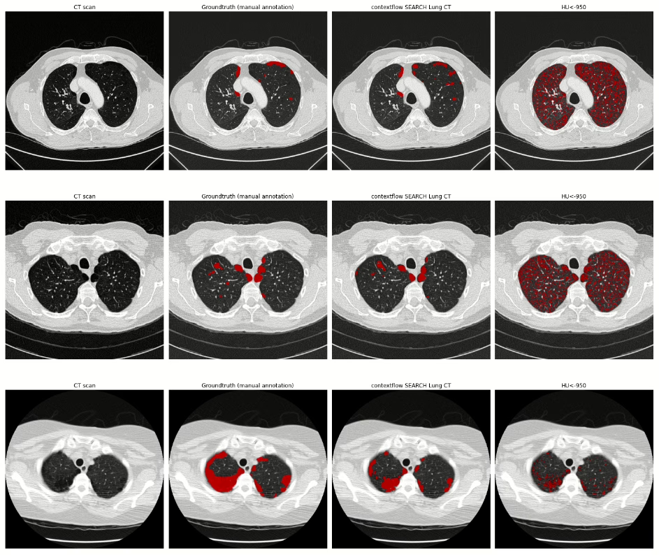
Using a threshold of -950 HU is a standard approach for quantification of emphysema in lung CT [Wang et al. 2013]. Although this thresholding method can be used to give an estimate of the emphysema volume, it is not suitable to identify the area of emphysema in detail. By contrast, contextflow SEARCH Lung CT utilizes a deep learning segmentation method to identify the exact area of emphysema and present more accurate volume measurements. When used for detection, the thresholding-based approach identifies emphysema areas in nearly all of the CT scans. In contrast, the AI-based approach has better specificity and thus reduces small false-positive occurrences.

We have assessed the performance of both methods (AI-based Emphysema segmentation by contextflow SEARCH Lung CT; standard HU-based thresholding, no cluster analysis) on 3617 slices drawn from a set of 494 scans with pixel-wise expert annotations. Ground truth was established through manual pixel-wise labeling of emphysematous image regions performed and included a quality control procedure involving expert radiologists.

Results are presented in table 1. The contextflow SEARCH method is more accurate in delineating emphysema locally on a pixel-level. The Dice metric is the standard way to assess the overlap of a segmentation method against the ground truth. The AI method achieves a Dice of 0.4 in contrast to a Dice of 0.23 with the thresholding-based method. Additionally, the volume of emphysema predicted in the slice is closer to the ground truth as can be seen from the volume similarity.

To assess emphysema detection on a case-level, we used a set of 494 positive and 559 negative scans (492 with other pathologies and 67 healthy cases). The contextflow SEARCH method has a similar sensitivity to the thresholding-based approach, detecting nearly every occurrence of the pattern. However the thresholding-based method predicts emphysema in every scan, resulting in 0% specificity. The AI-based method achieves a specificity of 56% and therefore reduces the overhead of false-positive occurrences presented to radiologists.

To better assess the impact of small emphysema predictions, e.g. due to noise in the image, we repeated the evaluation with different coverage cut-offs. Here, emphysema detection is only considered if at least 2%, 5%; or 10% respectively of the overall lung volume is covered with emphysema. Table 2 summarizes the corresponding findings, and shows that the overall difference remains consistent across these cut-offs.

Both datasets include scans with and without contrast enhancement, soft and sharp reconstruction kernels, and a slice thickness ranging from 0.75 to 5 mm. Varying acquisition parameters have been shown to influence the cutoff for emphysema detection [Boedeker et al. 2004, Gierada et al. 2010]; therefore the HU thresholding technique is expected to be more sensitive to the changes in acquisition than the method in contextflow SEARCH Lung CT.

The illustration below provides a visual comparison of results of both approaches for four chest CT scans. The first column (CT scan) shows the image data, the second column visualizes ground truth annotations for emphysema (manual pixel-wise annotations by expert annotators), the third column visualizes segmentation results provided by contextflow SEARCH Lung CT and the fourth column shows segmentation results for the HU-based thresholding approach.

Summary

Estimating the extent of emphysema based on an HU threshold was introduced in the 1990s [Coxson et al. 1990], [Gevenois et al. 1996]. With the recent progress in AI medical imaging, we are now able to measure the extent of emphysema more accurately, which is an important step towards detecting emphysema subtypes on a variety of lung CT scanner types. 

In combination with other COPD-relevant patterns [Lynch et al. 2015] that are also supported by contextflow SEARCH Lung CT, this is becoming the new basis for improved decision making in the treatment and progression of COPD patients.

If you have any questions, comments, are interested in a partnership or getting contextflow integrated into your clinical routine, feel free to contact us at office@contextflow.com.

References

Boedeker KL, McNitt-Gray MF, Rogers SR, Truong DA, Brown MS, Gjertson DW, Goldin JG. Emphysema: effect of reconstruction algorithm on CT imaging measures. Radiology. 2004 Jul;232(1):295-301.

Gierada DS, Bierhals AJ, Choong CK, Bartel ST, Ritter JH, Das NA, Hong C, Pilgram TK, Bae KT, Whiting BR, Woods JC. Effects of CT section thickness and reconstruction kernel on emphysema quantification: relationship to the magnitude of the CT emphysema index. Academic radiology. 2010 Feb 1;17(2):146-56.

Gevenois PA, De Vuyst P, de Maertelaer V, et al. Comparison of computed density and microscopic morphometry in pulmonary emphysema. Am J Respir Crit Care Med. 1996;154(1):187–192. doi:10.1164/ajrccm.154.1.8680679

Coxson HO, Rogers RM, Whittall KP, et al. A quantification of the lung surface area in emphysema using computed tomography. Am J Respir Crit Care Med. 1999;159(3):851–856. doi:10.1164/ajrccm.159.3.9805067

Sekine, Y., Katsura, H., Koh, E., Hiroshima, K. and Fujisawa, T., 2012. Early detection of COPD is important for lung cancer surveillance. European Respiratory Journal, 39(5), pp.1230-1240.

Lynch DA, Austin JH, Hogg JC, et al. CT-definable subtypes of chronic obstructive pulmonary disease: a statement of the Fleischner Society. Radiology. 2015;277(1):192–205. doi:10.1148/radiol.2015141579

Labaki, W.W. and Han, M.K., 2018. Improving detection of early chronic obstructive pulmonary disease. Annals of the American Thoracic Society, 15(Supplement 4), pp.S243-S248

González, G., Ash, S.Y., Vegas-Sánchez-Ferrero, G., Onieva Onieva, J., Rahaghi, F.N., Ross, J.C., Diaz, A., San José Estépar, R. and Washko, G.R., 2018. Disease staging and prognosis in smokers using deep learning in chest computed tomography. American journal of respiratory and critical care medicine, 197(2), pp.193-203.

Johnson, K.M., Sadatsafavi, M., Adibi, A., Lynd, L., Harrison, M., Tavakoli, H., Sin, D.D. and Bryan, S., 2021. Cost effectiveness of case detection strategies for the early detection of COPD. Applied Health Economics and Health Policy, 19(2), pp.203-215.

Wang, Z., Gu, S., Leader, J. K., Kundu, S., Tedrow, J. R., Sciurba, F. C., Gur, D., Siegfried, J. M., and Pu, J., 2013. Optimal threshold in CT quantification of emphysema. European radiology, 23(4), 975–984. https://doi.org/10.1007/s00330-012-2683-z

Other News

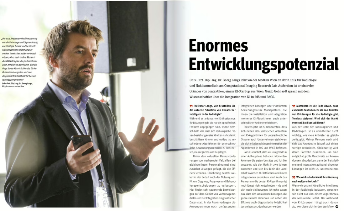
Enormous Development Potential
2022-12-21

How will the AI in radiology industry continue to evolve in the coming years? Will there be consolidation? What do radiologists need to be aware of when evaluating AI-based medical devices? Chief Scientist & Co-Founder Georg Langs was interviewed by Guido Gebhardt for Radiologie Magazine about these timely topics…and more. The full article can be found here. (German)

Other News

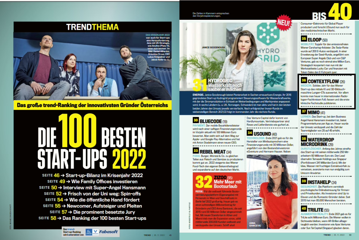
Trend.at places contextflow in their list of Top Austrian Startups
2022-12-21

For the fourth year in a row, contextflow has been listed in the Trend.at list of top 100 startups in Austria for 2022. This year, we placed 36! See all the worthy honorees here or visit trend.at.

Other News

contextflow @RSNA 2022
2022-11-22

We’re kicking off RSNA with a bang! THIRTEEN partners will be showcasting contextflow at their booths. See the list below for more details.

Other News

AI advances lung cancer diagnostics
2022-11-21

contextflow integrates additional features into ADVANCE Chest CT

Software is ever-evolving, as do user requirements. So it is only logical that companies benefit from the know-how of their customers. At least that’s how contextflow views the product development process. The Vienna-based machine learning in radiology expert has implemented corresponding suggestions into its new version of ADVANCE Chest CT.

Brand new for RSNA in Chicago is the name ADVANCE Chest CT. contextflow’s core product was previously named SEARCH Lung CT, but additional features necessitated an update in the product’s name, which also suggests an ADVANCEing forward of innovation to combat interstitial lung diseases, COPD and lung cancer. And these advances are paying off: contextflow will be shown or discussed at twelve partner booths at RSNA22!

One of ADVANCE Chest CT’s most-requested and newest features is TIMELINE for lung nodules. It automatically visualizes and quantifies changes in lung nodules over time, allowing radiologists to view multiple prior exams side-by-side. “Radiologists tell us they spend a lot of time preparing for follow-up examinations and tumor boards. With TIMELINE, they can view prior scans instantly, with consistent measurements of nodule characteristics, and we expect it will save radiologists a great deal of time,” says Marcel Wassink, Chief Commercial Officer at contextflow.

Another innovation, the integration of RevealAI-Lung from RevealDx into ADVANCE Chest CT, also supports the diagnosis of lung cancer by indicating malignancy similarity index for each nodule. “Lung cancer screening is currently expensive and slow, and it often leads to unnecessary procedures and stress for the patient. Implementing the lung nodule characterization component from RevealAI-Lung was a no-brainer because its clinical evaluation showed it could significantly impact on clinical decision making,” says contextflow Chief Product Officer Markus Krenn. In a clinical study published in September in the Journal of the American College of Radiology, it has been shown that the number of false positive and negative findings can be significantly reduced. When implemented in clinical routine, this could not only save resources, but also reduce stress for patients by avoiding unnecessary examinations.

In addition to RevealAI-Lung, Elsevier’s STATdx is integrated into ADVANCE Chest CT: STATdx provides radiologists with a list of possible differential diagnoses for a defined finding. With 1,400 differential diagnosis modules, the software includes more than 4,700 common and complex diagnoses with 200,000 image examples. “Through this partnership, we can support our users in faster and simpler reporting. In addition, Elsevier provides a systematic approach that allows radiologists to earn CME points virtually on the side,” says Marcel Wassink, describing the advantages of the integration.

For information on how contextflow ADVANCE Chest CT can support you with complex ILD, COPD and lung cancer cases, contact sales@contextflow.com or visit contextflow at RSNA: AI Showcase in South Hall Level 3, Booth 4649.

Other News

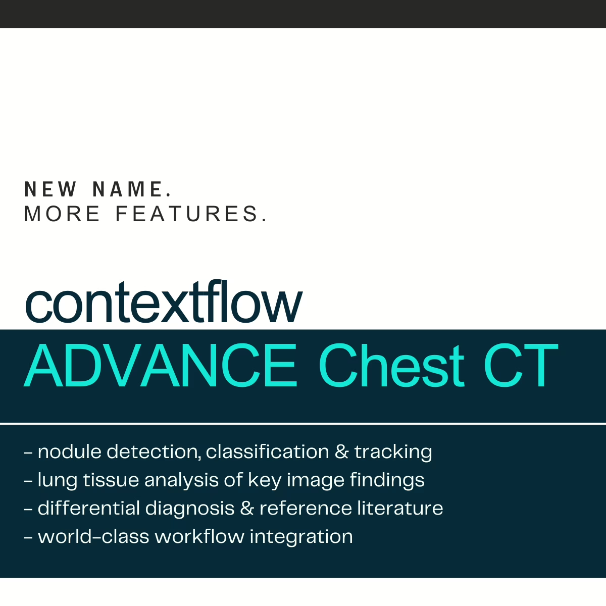
Out with the old, ADVANCEing towards the new! Announcing our new product name: contextflow ADVANCE Chest CT!
2022-10-28

Why? As our capabilities have grown, we recognize that SEARCH Lung CT no longer properly encompasses the wide range of features we now offer. What started as a 3D search engine for medical images now comprises a comprehensive computer-aided detection software for the entire chest, including quantitative information for suspected ILD, COPD and lung cancer cases. ADVANCE also reflects our constant push forward towards transparent, integrated AI for radiologists. 

What? contextflow ADVANCE Chest CT officially replaces the name SEARCH Lung CT. No worries – You can still access SEARCH as part of ADVANCE; only now you also get nodule detection, nodule tracking and lung tissue analysis all-in-one! And all integrated directly into your PACS, of course;)

When? Name changes take time, so we’re currently working on updating our website and materials to reflect this update.   

Other News