Medexprim, the European leader in multiomic real-world datasets for clinical research, is pleased to announce its partnership with contextflow, the Viennese-based medical device manufacturer known for its innovative computer-aided detection software for chest CT.
The Medexprim Suite™ extracts, aggregates, curates, enriches, and deidentifies imaging and clinical data to create regulatory-grade, multicentric, multiomic datasets for clinical research projects. The company provides its GDPR-compliant solution to a network of European university hospitals and cancer centres. Founded in 2016, contextflow offers comprehensive computer-aided detection support for ILD, COPD and lung cancer. Its core product, ADVANCE Chest CT, detects, quantifies, and visualises lung nodules and critical lung disease patterns.
In 2023 Medexprim will produce a European highly curated dataset of 3000 non-small cell lung cancer (NSCLC) cases with aggregated diagnostic and follow-up images contextualised with clinical data. contextflow ADVANCE Chest CT fully integrates into Medexprim Suite™ and is able to analyze this longitudinalcollection of CT scans, automatically label these images and indicate treatment efficacy by measuring disease progression over time.
The partnership will enable both the retrospective analysis of a patient images along with their profiles, treatment history, and treatment response. Future training of the model will allow for better diagnosis and prognosis.
Regarding the partnership, Romain Cazavan, CEO of Medexprim, says: “contextflow can be used in daily routine care to detect and measure lung nodules and many more patterns, but it also can be used longitudinally to better understand the progression of the disease compared to treatment. In the future, we would like to be able to predict potential disease regression and offer proactive advice for adapting treatment. Patient timeline is key, and our mutual expertise acts as a wonderful sandbox to streamline routine care.”
Marcel Wassink, CCO at contextflow, continues: “Innovation in medical diagnosis and care is often difficult and slow partly due to the lack of curated data. With this partnership with Medexprim, we are happy to contribute to the faster curation of large imaging data sets and help speed up the realization of new innovations in the market.”
This whitepaper was developed by contextflow’s Scientific and R&D teams to explain our approach behind emphysema quantification and why HU is not the best approach. The full text is listed below. For a pdf copy, click here.
Emphysema quantification with contextflow SEARCH Lung CT
Emphysema detection and volumetry in lung CT serves as an important factor in COPD detection and is relevant for timely, effective treatment and risk prediction for acute respiratory events [González et al. 2018], prognosis [Labaki and Han 2018], or lung cancer surveillance [Sekine et al. 2012]. Recent evidence shows that early COPD detection has clear advantages for patients and is cost effective [Johnson et al. 2021].
Using a threshold of -950 HU is a standard approach for quantification of emphysema in lung CT [Wang et al. 2013]. Although this thresholding method can be used to give an estimate of the emphysema volume, it is not suitable to identify the area of emphysema in detail. By contrast, contextflow SEARCH Lung CT utilizes a deep learning segmentation method to identify the exact area of emphysema and present more accurate volume measurements. When used for detection, the thresholding-based approach identifies emphysema areas in nearly all of the CT scans. In contrast, the AI-based approach has better specificity and thus reduces small false-positive occurrences.
We have assessed the performance of both methods (AI-based Emphysema segmentation by contextflow SEARCH Lung CT; standard HU-based thresholding, no cluster analysis) on 3617 slices drawn from a set of 494 scans with pixel-wise expert annotations. Ground truth was established through manual pixel-wise labeling of emphysematous image regions performed and included a quality control procedure involving expert radiologists.
Results are presented in table 1. The contextflow SEARCH method is more accurate in delineating emphysema locally on a pixel-level. The Dice metric is the standard way to assess the overlap of a segmentation method against the ground truth. The AI method achieves a Dice of 0.4 in contrast to a Dice of 0.23 with the thresholding-based method. Additionally, the volume of emphysema predicted in the slice is closer to the ground truth as can be seen from the volume similarity.
To assess emphysema detection on a case-level, we used a set of 494 positive and 559 negative scans (492 with other pathologies and 67 healthy cases). The contextflow SEARCH method has a similar sensitivity to the thresholding-based approach, detecting nearly every occurrence of the pattern. However the thresholding-based method predicts emphysema in every scan, resulting in 0% specificity. The AI-based method achieves a specificity of 56% and therefore reduces the overhead of false-positive occurrences presented to radiologists.
To better assess the impact of small emphysema predictions, e.g. due to noise in the image, we repeated the evaluation with different coverage cut-offs. Here, emphysema detection is only considered if at least 2%, 5%; or 10% respectively of the overall lung volume is covered with emphysema. Table 2 summarizes the corresponding findings, and shows that the overall difference remains consistent across these cut-offs.
Both datasets include scans with and without contrast enhancement, soft and sharp reconstruction kernels, and a slice thickness ranging from 0.75 to 5 mm. Varying acquisition parameters have been shown to influence the cutoff for emphysema detection [Boedeker et al. 2004, Gierada et al. 2010]; therefore the HU thresholding technique is expected to be more sensitive to the changes in acquisition than the method in contextflow SEARCH Lung CT.
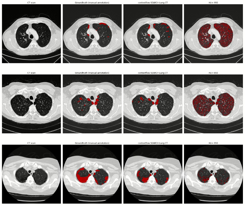
The illustration below provides a visual comparison of results of both approaches for four chest CT scans. The first column (CT scan) shows the image data, the second column visualizes ground truth annotations for emphysema (manual pixel-wise annotations by expert annotators), the third column visualizes segmentation results provided by contextflow SEARCH Lung CT and the fourth column shows segmentation results for the HU-based thresholding approach.
Summary
Estimating the extent of emphysema based on an HU threshold was introduced in the 1990s [Coxson et al. 1990], [Gevenois et al. 1996]. With the recent progress in AI medical imaging, we are now able to measure the extent of emphysema more accurately, which is an important step towards detecting emphysema subtypes on a variety of lung CT scanner types.
In combination with other COPD-relevant patterns [Lynch et al. 2015] that are also supported by contextflow SEARCH Lung CT, this is becoming the new basis for improved decision making in the treatment and progression of COPD patients.
If you have any questions, comments, are interested in a partnership or getting contextflow integrated into your clinical routine, feel free to contact us at office@contextflow.com.
References
Boedeker KL, McNitt-Gray MF, Rogers SR, Truong DA, Brown MS, Gjertson DW, Goldin JG. Emphysema: effect of reconstruction algorithm on CT imaging measures. Radiology. 2004 Jul;232(1):295-301.
Gierada DS, Bierhals AJ, Choong CK, Bartel ST, Ritter JH, Das NA, Hong C, Pilgram TK, Bae KT, Whiting BR, Woods JC. Effects of CT section thickness and reconstruction kernel on emphysema quantification: relationship to the magnitude of the CT emphysema index. Academic radiology. 2010 Feb 1;17(2):146-56.
Gevenois PA, De Vuyst P, de Maertelaer V, et al. Comparison of computed density and microscopic morphometry in pulmonary emphysema. Am J Respir Crit Care Med. 1996;154(1):187–192. doi:10.1164/ajrccm.154.1.8680679
Coxson HO, Rogers RM, Whittall KP, et al. A quantification of the lung surface area in emphysema using computed tomography. Am J Respir Crit Care Med. 1999;159(3):851–856. doi:10.1164/ajrccm.159.3.9805067
Sekine, Y., Katsura, H., Koh, E., Hiroshima, K. and Fujisawa, T., 2012. Early detection of COPD is important for lung cancer surveillance. European Respiratory Journal, 39(5), pp.1230-1240.
Lynch DA, Austin JH, Hogg JC, et al. CT-definable subtypes of chronic obstructive pulmonary disease: a statement of the Fleischner Society. Radiology. 2015;277(1):192–205. doi:10.1148/radiol.2015141579
Labaki, W.W. and Han, M.K., 2018. Improving detection of early chronic obstructive pulmonary disease. Annals of the American Thoracic Society, 15(Supplement 4), pp.S243-S248
González, G., Ash, S.Y., Vegas-Sánchez-Ferrero, G., Onieva Onieva, J., Rahaghi, F.N., Ross, J.C., Diaz, A., San José Estépar, R. and Washko, G.R., 2018. Disease staging and prognosis in smokers using deep learning in chest computed tomography. American journal of respiratory and critical care medicine, 197(2), pp.193-203.
Johnson, K.M., Sadatsafavi, M., Adibi, A., Lynd, L., Harrison, M., Tavakoli, H., Sin, D.D. and Bryan, S., 2021. Cost effectiveness of case detection strategies for the early detection of COPD. Applied Health Economics and Health Policy, 19(2), pp.203-215.
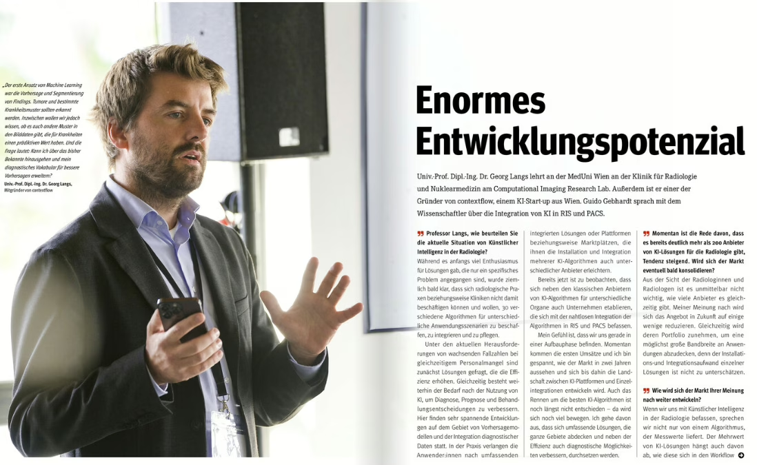
How will the AI in radiology industry continue to evolve in the coming years? Will there be consolidation? What do radiologists need to be aware of when evaluating AI-based medical devices? Chief Scientist & Co-Founder Georg Langs was interviewed by Guido Gebhardt for Radiologie Magazine about these timely topics…and more. The full article can be found here. (German)
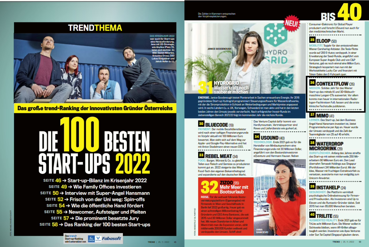
For the fourth year in a row, contextflow has been listed in the Trend.at list of top 100 startups in Austria for 2022. This year, we placed 36! See all the worthy honorees here or visit trend.at.
contextflow integrates additional features into ADVANCE Chest CT
Software is ever-evolving, as do user requirements. So it is only logical that companies benefit from the know-how of their customers. At least that’s how contextflow views the product development process. The Vienna-based machine learning in radiology expert has implemented corresponding suggestions into its new version of ADVANCE Chest CT.
Brand new for RSNA in Chicago is the name ADVANCE Chest CT. contextflow’s core product was previously named SEARCH Lung CT, but additional features necessitated an update in the product’s name, which also suggests an ADVANCEing forward of innovation to combat interstitial lung diseases, COPD and lung cancer. And these advances are paying off: contextflow will be shown or discussed at twelve partner booths at RSNA22!
One of ADVANCE Chest CT’s most-requested and newest features is TIMELINE for lung nodules. It automatically visualizes and quantifies changes in lung nodules over time, allowing radiologists to view multiple prior exams side-by-side. « Radiologists tell us they spend a lot of time preparing for follow-up examinations and tumor boards. With TIMELINE, they can view prior scans instantly, with consistent measurements of nodule characteristics, and we expect it will save radiologists a great deal of time, » says Marcel Wassink, Chief Commercial Officer at contextflow.
Another innovation, the integration of RevealAI-Lung from RevealDx into ADVANCE Chest CT, also supports the diagnosis of lung cancer by indicating malignancy similarity index for each nodule. « Lung cancer screening is currently expensive and slow, and it often leads to unnecessary procedures and stress for the patient. Implementing the lung nodule characterization component from RevealAI-Lung was a no-brainer because its clinical evaluation showed it could significantly impact on clinical decision making, » says contextflow Chief Product Officer Markus Krenn. In a clinical study published in September in the Journal of the American College of Radiology, it has been shown that the number of false positive and negative findings can be significantly reduced. When implemented in clinical routine, this could not only save resources, but also reduce stress for patients by avoiding unnecessary examinations.
In addition to RevealAI-Lung, Elsevier’s STATdx is integrated into ADVANCE Chest CT: STATdx provides radiologists with a list of possible differential diagnoses for a defined finding. With 1,400 differential diagnosis modules, the software includes more than 4,700 common and complex diagnoses with 200,000 image examples. « Through this partnership, we can support our users in faster and simpler reporting. In addition, Elsevier provides a systematic approach that allows radiologists to earn CME points virtually on the side, » says Marcel Wassink, describing the advantages of the integration.
For information on how contextflow ADVANCE Chest CT can support you with complex ILD, COPD and lung cancer cases, contact sales@contextflow.com or visit contextflow at RSNA: AI Showcase in South Hall Level 3, Booth 4649.
Why? As our capabilities have grown, we recognize that SEARCH Lung CT no longer properly encompasses the wide range of features we now offer. What started as a 3D search engine for medical images now comprises a comprehensive computer-aided detection software for the entire chest, including quantitative information for suspected ILD, COPD and lung cancer cases. ADVANCE also reflects our constant push forward towards transparent, integrated AI for radiologists.
What? contextflow ADVANCE Chest CT officially replaces the name SEARCH Lung CT. No worries – You can still access SEARCH as part of ADVANCE; only now you also get nodule detection, nodule tracking and lung tissue analysis all-in-one! And all integrated directly into your PACS, of course;)
When? Name changes take time, so we’re currently working on updating our website and materials to reflect this update.
Signify Research is widely-respected in our industry for its in-depth analyses of current market trends. That’s why we’re honored to have been featured in the article titled « contextflow Reveals Its Hand« , discussing the why behind our recent partnership announcment with RevealDx.
RevealDx developed RevealAIöLung, the world’s first CADx software for the characterization of lung nodules to receive the CE Mark. The company recently completed its pivotal clinical trial which demonstrated significant improvement in both early detection of malignant nodules and reduction in false positive nodules in both screening and incidental cohorts. By integrating this patented technology into clinical routine, healthcare providers can reduce unnecessary procedures, cost and stress for patients with lung nodules.
Signify Research’s take? « contextflow’s SEARCH solution remains unique in the market today. No other vendor offers a similar value proposition. Further, a study published in the Journal of the American College of Radiology highlighted Reveal DX’s ability to help clinicians diagnose lung cancer sooner with improved specificity. By bringing these capabilities together, both vendors have created a comprehensive lung cancer solution for CT imaging that can, on paper, outcompete most of its competitors. »
RevealDx appoints contextflow as its exclusive distributor in the European Union and selected territories. The companies also announce trial installation discounts for select customers.
Chest specialists RevealDx and contextflow announced today an agreement for exclusive distribution of RevealDx’ products in the EU and other selected territories. The companies will be launching their integrated solution at the upcoming IASLC World Conference on Lung Cancer in Vienna in August.
RevealDx developed RevealAI-Lung, the world’s first CADx software for the characterization of lung nodules to receive the CE Mark. The company recently completed its pivotal clinical trial which demonstrated significant improvement in both early detection of malignant nodules and reduction in false positive nodules in both screening and incidental cohorts.* By integrating this patented technology into clinical routine, healthcare providers can reduce unnecessary procedures, cost and stress for patients with lung nodules. *publication Fall 2022
contextflow is an industry leader in AI-based medical devices for radiologists evaluating chest CTs. Its core technology is SEARCH Lung CT, a clinical decision support system that automatically detects, quantifies and visualizes key disease patterns and lung nodules in CTs of the lungs over time, displaying relevant information directly in the radiologist’s PACS viewer. The tool is relevant for the analysis of many suspected diseases, including interstitial lung disease (ILD), chronic obstructive pulmonary disease (COPD) and lung cancer. By including RevealDx’ lung nodule characterization technology into SEARCH Lung CT, contextflow strengthens its capabilities even further in the area of lung cancer screening by providing not just quantifications but also classification of lung nodule types.
Integrating RevealDx’ nodule characterization technology into SEARCH Lung CT was a logical next step for both sides. As Chris Wood, CEO of RevealDx says, “We are thrilled to be partnering with contextflow. They have developed the most comprehensive set of tools for reading chest CT available today. Adding RevealAI-Lung to their system makes it far and away the best solution to efficiently and accurately interpret these challenging lung cancer cases.”
Markus Holzer, CEO of contextflow continues, “The RevealAI-Lung software has been well integrated into our software, creating a seamless experience for users. We recently launched our nodule detection software and quickly realized that characterization of nodules is essential to make a significant impact on patient care. Partnering with RevealDx adds a layer of detail that we feel will become indispensable for reducing false positives and false negatives during lung cancer screening as well as in standard clinical routine.”
To schedule a virtual demo or book an appointment at IASLC, contact sales@contextflow.com.
About RevealDx
RevealDx developed RevealAI-Lung, the world’s first CADx software for the characterization of lung nodules to receive the CE Mark. RevealAI-Lung has been validated in clinical studies that show improvement in diagnostic precision using our patented methods. Results demonstrate the software can significantly accelerate lung cancer diagnosis and reduce unnecessary procedures. https://reveal-dx.com/
Our first clinical study in the European Radiology Journal from the European Society of Radiology has been published! The study, in collaboration with the Medizinische Universität Wien and Universitätsklinikum AKH Wien shows reading time decreased by 31% when contextflow SEARCH Lung CT was available for use.