
The value of AI is in its integration – 3 Pioneers from LUMC share their experiences using contextflow integrated into the Sectra PACS
2022-07-14

Sectra interviewed three thought leaders from Leiden University Medical Center (LUMC) in the Netherlands about their experiences and insights on integrating AI directly into the radiology workflow. This use case shows the importance of that integration, how it’s used in clinical practice, and how they went about choosing the right types of algorithms for the hospital. 

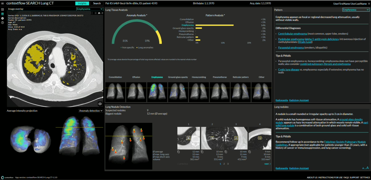
LUMC uses contextflow’s clinical decision support system for improved analysis of lung CTs to enhance its radiology workflow with artificial intelligence. The software, SEARCH Lung CT, has been seamlessly integrated into the Sectra PACS, enabling radiologists to quantify lung abnormalities, such as lung nodules, emphysema, effusion, consolidation, pneumothorax, and many more. The software serves to assist radiologists with both quantitative and qualitative information when evaluating patients with suspected interstitial lung diseases (ILD), chronic obstructive pulmonary disease (COPD), and lung cancer. Using this AI assistance, it may also contribute to the wellbeing of radiologists at work, thus going beyond work efficiency and quality enhancement of radiological reporting.

  • Prof. Dr. H. Lamb, Professor of Radiology, Head of the Cardio Vascular Imaging Group (CVIG), Department of Radiology
  • Dr. W. Grootjans, Technical Physician, Head of the Imaging Services Group, Department of Radiology
  • S. Romeijn, Technical Physician, clinical implementation of AI, Department of Radiology

Trends in radiology: more data, higher workloads, better PACS integrations

In recent years, data volumes in radiology have increased dramatically. “We have an ever growing number of clinical imaging requests. Exams have more slices and require a higher resolution, so there is much more imaging data to review and analyze quantitatively,” says professor H. Lamb. Dr. W. Grootjans adds: “Image quantification has become a standard task in radiology, and its importance will only continue to increase. It provides referring physicians with the necessary information to personalize patient care, improve sensitivity to detect changes over time, which ultimately aims to improve patient outcomes.”

In addition, general awareness of the potential of AI is increasing. Lamb: “Since the radiological workload continues to increase and the number of radiologists will not increase, we have a challenge. AI will help to maintain high quality of image reading with sustainable well-being of radiologists, allowing them to focus more on their role as consultant and communicator. Radiology with AI support is shifting the radiologist’s role towards a navigator of healthcare. We need to keep radiologists satisfied in their jobs and likewise offer our patients good turnaround times.” 

Numerous AI algorithms have been developed in the past few years. This brings plenty of choice, but working with AI goes beyond choosing to work with a specific tool. S. Romeijn explains: “Algorithms often perform very well, but that is only the first step. The second step is integration because you can’t do anything without this. Most of the added value of AI is in its optimal software integration, in this case the PACS, which helps us get something out of it in practice. For radiologists, the right data must be in the right place at the right time. This also stimulates radiologists’ enthusiasm.” 

Choosing to work with contextflow

Dozens of companies worldwide are developing algorithms to detect lung abnormalities. LUMC chose contextflow SEARCH Lung CT, which is now one of the first tools that LUMC has integrated into its Sectra PACS. One deciding factor was this developer’s openness to improvement. “To us, the [nature of our] collaboration is an essential factor, and with this, their openness to change. You don’t just want to buy a license. You want them to customize their product and work on projects together. We are not simply their client, but we will also have to get along well with each other,” according to Lamb. 

The functionality of contextflow stood out, as it takes a general approach to image analysis. Romeijn: “Ultimately, contextflow is more widely applicable than other algorithms focusing on specific lung abnormalities. It’s a tool for the lungs and, therefore, a lot more interesting than having eight different tools that say something about a thorax CT, with all kinds of different outputs that are difficult to combine. So the vision of contextflow is very appealing to us.” 

PACS integration of contextflow and its improvements

The LUMC team met contextflow in 2018. “In those four years, we had a lot of meetings that set direction for the future. Ultimately, it’s about being able to implement your vision into the software,” says Grootjans. The integration of contextflow into the Sectra PACS started in 2020. “At the time, we were still exploring what AI was all about. Since it was one of the first AI algorithms to be implemented, we spent approximately one year on discussing IT security and filling in the paperwork. Then COVID-19 came. [At that point,] we were in touch with both Sectra and contextflow. An important thing to note is that this degree of AI integration into the PACS was unheard of before Sectra made it possible. Sectra is also currently the only PACS that is technically capable of integrating that deeply. On top of that, contextflow is one of the very few AI vendors capable of such a deep integration.”

Because it was a pioneering collaboration, all parties had to gain more insight, which then had to guide the workflow setup in the best possible way. Romeijn: “We catch up with contextflow every month. During the integration, this was daily or weekly via email to discuss the necessary tweaks and adjustments. They respond very quickly and take home our feedback.”

Formally established collaboration contracts define what the LUMC team provides and what contextflow will offer in return. “You could call these co-creation contracts,” Lamb explains. “We help them annotate lung abnormalities, and they create tailored solutions. You can’t do this from the start. First, you have to get to know and trust each other. Nowadays, we are at that stage where we can basically [try anything].” LUMC’s image annotations help validate the algorithms. In addition, the LUMC team can share their practical experience, from which contextflow learns what works well and what does not. This input will further determine the workflow and how the radiologist can interact with it.

At LUMC, the arrival of COVID-19 accelerated their pioneering with AI. Lamb: “Everyone had to work from home. Suddenly, we could start doing things we wanted to do for years. There was also a huge need for the quantification of lung patterns. What percentage of the lung was affected by COVID-19?” Many IT problems were solved during this period, including legislation-related ones.  

The integration of contextflow SEARCH Lung CT was a step-by-step process. Romeijn: “At first, we had to open a link in our PACS, directing us to a separate contextflow viewer. That was a nice integration by itself.” But the wish of the end users remained to have as much integration with the Sectra PACS as possible.   

Another improvement in the contextflow – Sectra integration is how its output can be transferred to reports. Lamb: “I [previously] couldn’t transfer the results from the analysis directly into my report. No software had this possibility. So we all asked for that.” Now it’s more a matter of checking and accepting the output before it is added to a report. This also prevents errors.

The application itself is also constantly improving. “We know that they thought carefully about longitudinal analyses. This feature has not yet been implemented, but it’s coming after this summer,” Romeijn explains. From then on, contextflow can also be used to properly visualize and quantify the development of lung abnormalities over time. Lamb: “This is what we told them from the beginning. We need to follow lung nodules over time, visualize them using graphs, doubling times, and more. They have developed this feature exactly as we want it in daily use. This development is truly a win-win. Collaboration offers you the best solutions.” According to Romeijn, it can be a challenge to test what works best in clinical practice. For example; how to choose which previous scans and series should be compared with the latest scan.

The impact of working with AI

What impact does working with AI have on radiologists? Lamb says this is difficult to say, “because how do you measure this impact?” In the Radiology AI Lab, he and his team are developing a method to quantify how focused radiologists stay in their tasks. “As radiologists, we take very few breaks. We sometimes start at 8 AM and go home at 8 PM, which shouldn’t be possible. We want to discover the parameters that are most informative about your reliability, efficiency, but above all, your job satisfaction.” This shows that working with tools like contextflow goes far beyond saving time and improving quality of care, but also about job satisfaction and preventing burnout.

Patient communication remains essential when working with AI. “They have access to their EHR (Electronic Health Record) data, but the wording is often very technical and medical. People can start panicking. We want to take that into account by having a simple patient explanation in the future,” Lamb says. Romeijn adds that if there is a difference between the AI output and what the radiologist sees, the radiologist should let the patient know why the difference exists and what findings prevail. 

At LUMC, referring clinicians have rapidly become used to the radiological output with the help of AI. Lamb explains that they see the luxury of it. “Even though it has taken some time to set up, it is a very efficient, quantitative way of communicating.” According to Grootjans, it is “vital to further involve the referring clinicians so that you keep looking at the workflow holistically. What information are you providing, and what is necessary?” Of course, the human factor will always be needed. Lamb: “I am not worried about that at all. I hope that everything will be automated at some point and that radiologists will translate this into clinical practice. They will become [more like] imaging consultants and can guide patients toward their next steps.”

Getting started with AI

As pioneers in applying AI in medical imaging, Lamb, Grootjans, and Romeijn continuously look ahead. First, portfolio management is important for every hospital. “Every hospital has a different [medical] portfolio. This portfolio selection informs the choice of algorithms you want to work with as a hospital,” says Grootjans.

Lamb recommends hospitals that start with AI to “make sure you integrate your radiology workflow in your PACS from the beginning. This helps you stay close to the images and data. The more technical aspects are complex, so you should maintain an excellent collaboration with your IT department.” 

He also emphasizes the importance of intermediaries with a background in technical medicine. In this way, the IT department better understands what the radiologists exactly need. For the LUMC, having technical physicians available to bridge implementation gap accelerated the adoption of AI in clinical practice.”
Other News

Chest experts Oxipit and contextflow team up for diagnostic quality assurance
2022-07-13

The new partnership aims to mitigate the risk associated with missed findings in CT medical imaging studies. The collaboration will combine Oxipit’s ChestEye Quality and contextflow’s SEARCH Lung CT products to identify missed findings in CT scans in near-real time. ECR 2022 will offer the first preview of the combined solution, and the first installation will be deployed at Leiden University Medical Center. 

ChestEye Quality analyzes medical images and corresponding radiologist reports. Acting as a virtual safety net, the software sends a notification to the radiologist if it detects a mismatch or a missed finding not identified in the radiologist report. 

ChestEye Quality can operate in retrospective and prospective settings, providing quality audit notifications in near-real time. The product is already deployed in more than 10 medical institutions worldwide. 

Out of nearly 200,000 analyzed chest X-ray images, an average of 1 in 552 (0.18%) chest X-ray studies feature clinically-significant missed findings. The result varies from 0.08% to 0.92% depending on the type of medical institution. 78% of the missed findings relate to pulmonary nodules, aiding earlier detection of lung cancer and significantly improving patient treatment prognosis.

The contextflow partnership will expand ChestEye Quality capabilities into the CT modality.

contextflow SEARCH Lung CT is a clinical decision support system that automatically detects, quantifies and visualizes key disease patterns and lung nodules in CTs of the lungs over time, displaying relevant information directly in the radiologist’s PACS viewer. The tool is relevant for the analysis of many suspected diseases, including interstitial lung disease (ILD), chronic pulmonary obstructive disease (COPD), and lung cancer.

In a clinical impact study at the Medical University of Vienna (MUW), an earlier version of SEARCH Lung CT showed an average reading time savings of 31% when contextflow SEARCH Lung CT is available for use with a trend towards improved diagnostic accuracy. The study was recently published in European Radiology.

“We are excited to partner with experts in CT AI medical imaging. The ChestEye Quality AI double reading approach has already proven itself in the CXR modality, helping radiologists to spot more clinically-relevant nodules and improving early diagnostics of lung cancer. Collaboration with contextflow highlights the robustness of the ChestEye Quality framework, showcasing how the AI double reader approach can be easily expanded into other medical imaging modalities,” says Oxipit CEO Gediminas Peksys.

contextflow Chief Commercial Officer Marcel Wassink continues: “Radiologists tell me they are often extremely busy or even exhausted towards the end of their shift. Reading lung CTs is a complicated task, whereby even the most experienced radiologists have only moderate consensus. Therefore they can’t deny they may sometimes oversee early signs of a disease in the scan or oversee or misjudge relevant patterns, which is supported by scientific publications. With this cooperation we aim to provide radiologists a safety net that catches potential mismatches between the contents in the radiology report and the visual findings related to all patterns in the CT scan detected by contextflow. The goal is to further support radiologists with a friendly warning system that helps them double check their analysis of the CT scan.”

The first ChestEye CT Quality deployment is planned at the Leiden University Medical Center (LUMC). 

“In the domain of chest X-ray and CT imaging, we have been successfully working with both Oxipit and contextflow for several years, with their applications integrated in the radiology workflow and in use in daily clinical practice. We are looking forward to having the quality functionality expanded to cover chest CT imaging with the goal of further improving the quality of care for our patients,” notes Head of Imaging Services Group at LUMC Dr Willem Grootjans. 

About Oxipit | www.oxipit.ai 

Oxipit develops AI applications for diagnostic medical imaging. With a team of award-winning data scientists and medical doctors, the company aims to introduce innovative artificial intelligence breakthroughs to everyday clinical practice.

About contextflow | www.contextflow.com 

contextflow is a spin-off of the Medical University of Vienna (MUW) and European research project KHRESMOI, supported by the Technical University of Vienna (TU). Founded by a team of AI and engineering experts in July 2016, the company has received numerous awards; most recently, contextflow was named a Born Global Champion 2021 by the Austrian Chamber of Commerce. Its clinical decision support software SEARCH Lung CT is CE Marked and available for clinical use within Europe under the new MDR.

Other News

STATdx and contextflow announce partnership to provide radiologists with improved tools to tackle differential diagnoses
2022-07-05
  • – New partnership provides improved clinical decision support for radiologists evaluating lung CTs
  • – Allows radiologists to earn Continuing Medical Education (CME) credits when exploring STATdx content from within contextflow SEARCH Lung CT

Elsevier is delighted to announce a new product integration combining Elsevier’s STATdx platform with the power of AI from contextflow SEARCH Lung CT. 

The integration supports radiologists when evaluating complex cases, saving time, increasing confidence and fostering ongoing learning for radiologists by allowing an automatic accumulation of CME credits. 

Elsevier’s STATdx platform enables users to gain expert diagnostic support, increasing speed, accuracy and confidence when reporting on a wide range of imaging. The contextflow SEARCH Lung CT system provides objective, qualitative and quantitative information for interstitial lung disease, chronic obstructive pulmonary disease (COPD) and lung cancer cases directly within the picture archiving and communications system (PACS). The integration of the two provides radiologists with the opportunity to expand their diagnostic support.

Dr. Kotter, contextflow advisor and Consultant in Diagnostic and Interventional Radiology, University of Freiburg – Medical Center outlined the benefits of the integration for radiologists, “Reporting differential diagnoses is a key task for radiologists. The combination of STATdx trusted content integrated into contextflow SEARCH Lung CT means that the radiologist has access to the best qualitative and quantitative information at their fingertips to report on complex cases. We have trialled this solution in our clinic, and we are very much looking forward to having this integration in place to support our daily clinical work.”  

Tim Morris, VP, GTM, EMEALAAP at Elsevier continued, “We are delighted to announce this new integration with contextflow, which provides radiologists with crucial support in their diagnoses. The combination of day-to-day clinical functionality whilst also fostering ongoing learning is an incredibly powerful tool for radiologists.”            

Marcel Wassink, Chief Commercial Officer at contextflow commented, “We are excited with this integration. Providing relevant differential diagnosis literature from STATdx directly to radiologists using contextflow will help them report chest CT cases faster and with more ease, whilst also accumulating CME credits.”

About Elsevier 

Elsevier is a global information analytics business that helps institutions and professionals progress science, advance healthcare and improve performance for the benefit of humanity. Elsevier provides digital solutions and tools in the areas of strategic research management, R&D performance, clinical decision support, and professional education; including ScienceDirect, Scopus, Scival, ClinicalKey and Sherpath. Elsevier publishes over 2,500 digitised journals, including The Lancet and Cell, more than 35,000 e-book titles and many iconic reference works, including Gray’s Anatomy. Elsevier is part of RELX Group, a global provider of information and analytics for professionals and business customers across industries. www.elsevier.com

Other News

contextflow launches a new version of SEARCH Lung CT
2022-03-22

Thanks to radiologists’ feedback, the latest version of SEARCH Lung CT now includes 3 new developments: 1) consolidation pattern is included as part of the quantitative image analysis results in the Insights Screen; 2) disease patterns can be shown color-coded in your viewer using DICOM secondary capture, 3) nodule detection results can be provided as a TID1500-compliant DICOM SR object.

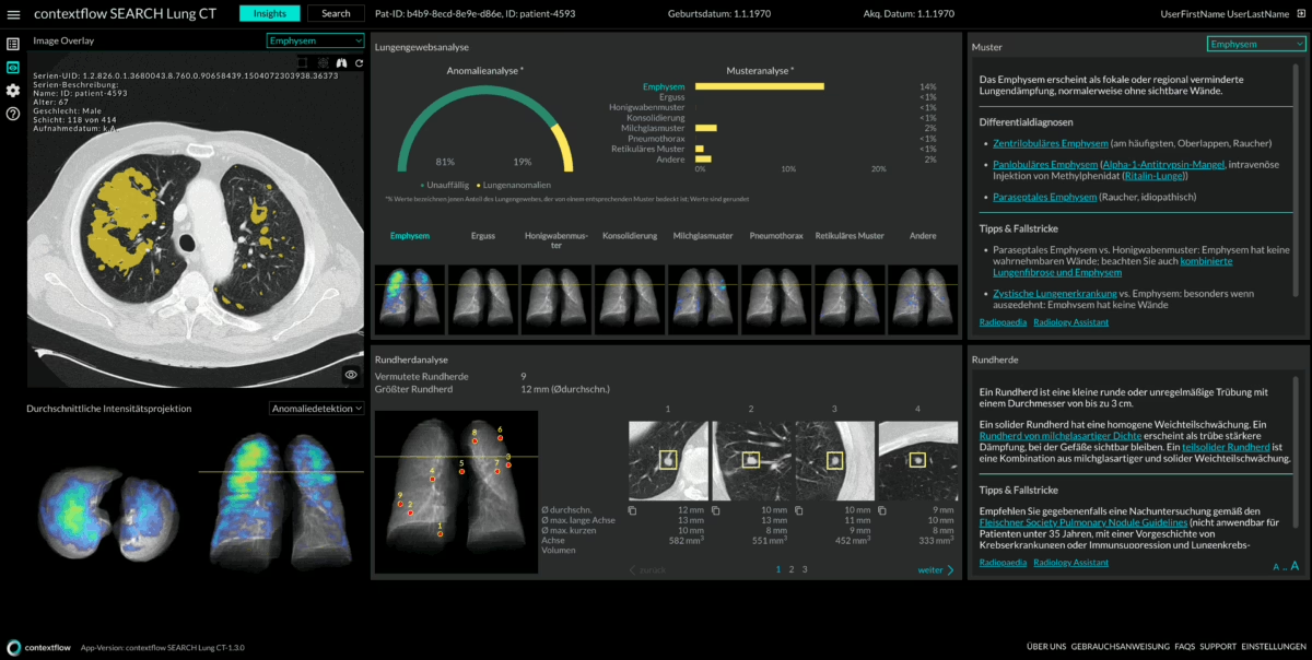
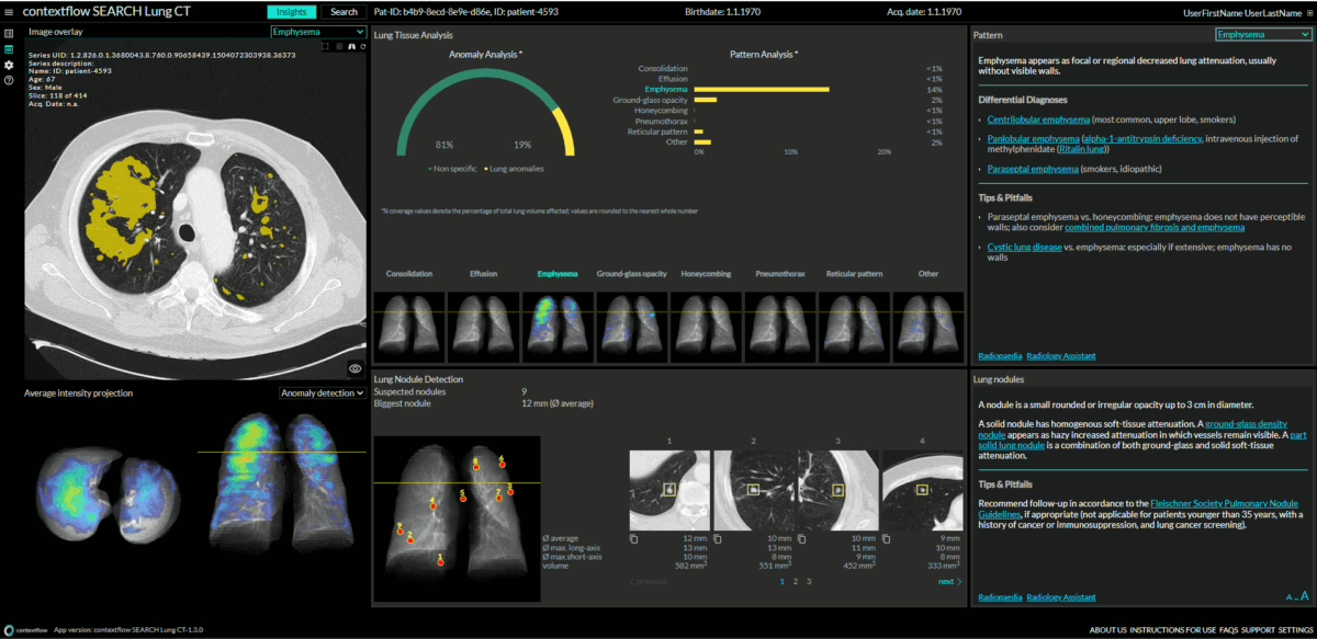
1) The Insights Screen now provides lung coverage values and distribution maps for 7 image patterns + visualization and measurements of detected lung nodules (top center above). The image patterns include: consolidation, effusion, emphysema, ground-glass opacity, honeycombing, pneumothorax, and reticular pattern.

2) These same 7 image patterns can be seen color-coded in your viewer using DICOM secondary capture…no need to click outof your viewer! (see below)

3) SEARCH Lung CT also provides nodule detection results in form of a DICOM Enhanced Structured Report object that follows DICOM Structured Reporting template TID1500.

The DICOM SR object lists all detected pulmonary nodules and provides the following information for those: location reference, long-axis diameter, short-axis diameter, average diameter, and volume.

The object is intended to be sent to and parsed by structured reporting systems or PACS for seamless integration of nodule detection results into the radiologists’ reporting workflow. If supported by your PACS, this feature allows you to accept/reject contextflow’s nodule results within your viewer.

For more information or to schedule a personalized demo, click here.

Other News

Oncological imaging – AI is indispensable for lung tissue classification
2022-06-28

Evangelisches Klinikum Niederrhein uses SEARCH Lung CT for distribution and volumetric quantifications of disease patterns

Has artificial intelligence (AI) come to stay? The answer is clearly yes, because from laboratory medicine to radiology, it is helping medical professionals obtain plausible, quantifiable and reproducible results in significantly less time. And as a result, it is easing the burden on medical departments suffering from large workloads. AI will actually take over many activities in the future in areas where it is readily applicable. “Repetitive work in our specialties, such as determining and matching values, is an excellent domain for the use of machines,” says Prof. Dr. Jörg Michael Neuerburg, Chief Physician of the Central Department of Diagnostic and Interventional Radiology at the Evangelisches Klinikum Niederrhein (EvKIN) Ev. Hospital BETHESDA.

The network is an academic teaching hospital, part of the University of Düsseldorf, and has five locations in the Ruhr region, including Ev. Klinikum Duisburg-Nord, Johanniter Krankenhaus Oberhausen, Herzzentrum Duisburg-Meiderich, Ev. Klinikum Dinslaken and Ev. Krankenhaus BETHESDA zu Duisburg. As a maximum care provider, the hospital operates a thorax center with two pulmonology departments as well as a heart center. The radiology department employs 16 radiologists and 9 neuroradiologists, serving half a million people from the Lower Rhine to the Ruhr region.

Artificial intelligence must be embedded

For AI to be accepted, reproducibility of results is an important factor; according to Prof. Neuerburg; however, complete integration into the usual radiological workflow is an absolute must. The problem with AI systems, as well as their predecessors – CAD systems, has always been implementation into the workflow: “If a separate program has to be opened and the images also have to be sent to another computer, the workflow is delayed. Radiology, like all other departments, is measured by throughput. If AI means additional work, acceptance is low. This problem has been solved very well by contextflow in collaboration with VISUS; SEARCH Lung CT is perfectly integrated into our workflow,” says Prof. Neuerburg.

The introduction of AI as a joint task

The radiologists at EvKIN work with the JiveX PACS from VISUS (Compugroup) and the ORBIS hospital information system from Dedalus HealthCare, both of which are already closely integrated. This provided good conditions for installing SEARCH Lung CT from contextflow in coordination with the pulmonologists. After the initial problems on the part of the legal department regarding data transfer were solved (it had to be ensured that no data protection guidelines would be affected during data transfer to other servers), the integration of the new program succeeded very quickly and without affecting ongoing operations thanks to the cooperation of the in-house IT, VISUS and contextflow.

The pitfalls of oncological imaging

In radiology, oncological imaging is a never-ending challenge: the targeted search for the tumor, its standardized classification, in the case of treatment, the assessment of its progress (or not). “Let’s take a patient with a large brain hemorrhage as an example. To answer the question of whether tumors were already present, the current examination must be compared with the previous examination. Of course, artificial intelligence can absolutely answer that much faster and more efficiently than we can,” says Prof. Neuerburg, describing a current case for the use of AI.

The chief radiologist already has broad experience with AI systems and uses three programs in his department: BoneXpert, an AI-based bone age determination system used in pediatrics to identify growth retardation or acceleration, and in forensics to determine the age of delinquent juveniles. Further AI support is provided by Transpara – a mammography screening tool that uses a graduation from zero to ten to indicate the probability of developing breast cancer.

For lung diagnostics, radiologists at EvKIN have been relying on SEARCH Lung CT for the past year to improve the overall quality and quantity of lung diagnostics, and in particular, to assist in assessing the distribution pattern of emphysema. “These distribution patterns are important to pulmonologists because they serve as the basis for setting valves to adequately ventilate the lungs. Therefore, we have adapted our findings to provide quantitative results on the extent to which, for example, the upper lobe is ventilated differently than the middle lobe after valve placement,” the radiologist explains.

In addition, SEARCH Lung CT is used for lung nodule detection staging during follow-ups. Previous examinations are compared with the current results to identify increasing structural densities. In addition, the system detects new nodules and measures the volumes of the existing ones; thus, it enables an assessment of a treatment’s progress. Thus, the radiologists at EvKIN mainly rely on quantitatively measurable changes. “SEARCH Lung CT is currently still in the development phase, so we use the tool as an add-on and report the volume information as a supplement to our findings without these values being standardized in the workflow,” explains Prof. Neuerburg.

On the expert’s wishlist is the expansion of the software to include the pleural region, for example, to detect occupational diseases such as mesothelioma, which occurs after exposure to asbestos. At the moment, the tool analyzes the lungs, but not all structures. Therefore, the radiologist’s additional visual findings are still necessary at this stage, especially since in the case of bronchial carcinomas, a look at the adrenal glands or the liver is also advisable to see whether metastases of the primary tumor have formed in the abdomen.

Standardization and classification pave the way for AI 

After a long lead-up, radiology is now moving swiftly toward standardized findings. What began years ago with the BI-RADS classifications in breast cancer screening has now become established via the PI-RADS classification in prostate imaging and the LI-RADS and ACR classifications in lung screening: a stringent and institution-independent staging system. This refers to the classification of tumors into specific disease stages, which subsequently require different treatment. For example, metastasized diseases are not only removed surgically; whereas early stages related to the organ can certainly be treated surgically, depending on the histology. “This is the direction in which AI will develop and have a significant impact on the lives of radiologists in the future. Because this is where the volumetry of lesions and volumetric comparison come into play, which is now mandatory for tumor center certification as part of oncology standardized staging.

“On the way to standardized reporting in radiology, the software from contextflow will therefore provide us with important support,” concludes Prof. Neuerburg, hinting at the latest paths in medical imaging.

Other News

Onkologische Bildgebung – KI zur Lungengewebeklassifizierung unverzichtbar
2022-06-28

Ev. Klinikum Niederrhein nutzt SEARCH Lung CT für volumetrische Bestimmungen und Übersicht über Verteilungsmuster

Ist die Künstliche Intelligenz (KI) gekommen, um zu bleiben? Die Antwort lautet eindeutig ja, denn von der Labormedizin bis zur Radiologie unterstützt sie Mediziner dabei, plausible, quantifizierbare und reproduzierbare Ergebnisse in deutlich kürzerer Zeit zu erhalten. Damit entlastet sie die unter großem Arbeitsaufkommen leidenden medizinischen Abteilungen. KI wird viele Tätigkeiten in den Bereichen, wo sie gut einsetzbar ist, künftig auch tatsächlich übernehmen. „Sich wiederholende Arbeiten in unseren Fachgebieten wie beispielsweise Werte zu ermitteln und abzugleichen, sind eine hervorragende Domäne für den Einsatz von Maschinen“, ist Prof. Dr. Jörg Michael Neuerburg, Chefarzt der zentralen Abteilung für Diagnostische und Interventionelle Radiologie, Evangelisches Klinikum Niederrhein (EvKlN) Ev. Krankenhaus BETHESDA, überzeugt.

Der Verbund ist Akademisches Lehrkrankenhaus der Universität Düsseldorf und hat mit dem Ev. Klinikum Duisburg-Nord, dem Johanniter Krankenhaus Oberhausen, dem Herzzentrum Duisburg-Meiderich, dem Ev. Krankenhaus Dinslaken und dem Ev. Krankenhaus BETHESDA zu Duisburg fünf Standorte im Ruhrgebiet. Als Maximalversorger betreibt das Klinikum ein Thoraxzentrum mit zwei pulmologischen Abteilungen sowie ein Herzzentrum. In der radiologischen Abteilung arbeiten 16 Radiologen und 9 Neuroradiologen, die ein Einzugsgebiet von einer halben Million Menschen vom Niederrhein bis zum Ruhrgebiet betreuen.

Künstliche Intelligenz muss eingebettet sein

Für die Akzeptanz beim Einsatz von KI ist nicht nur die Reproduzierbarkeit der Ergebnisse ein wichtiger Faktor, sondern laut Prof. Dr. Neuerburg auch die vollständige Integration in den gewohnten radiologischen Workflow ein absolutes Muss. Das Problem der KI-Systeme wie auch ihrer Vorläufer, den CAD-Systemen, war immer die Implementierung in den Arbeitsablauf: „Wenn ein separates Programm geöffnet werden muss und die Bilder möglicherweise zusätzlich an einen anderen Rechner geschickt werden müssen, verzögert sich der Arbeitsablauf. Die Radiologie, wie auch alle anderen Abteilungen, wird nach Durchsatz bemessen. Wenn KI zusätzliche Arbeit bedeutet, ist die Akzeptanz gering. Dieses Problem hat contextflow in Zusammenarbeit mit VISUS sehr gut gelöst,      SEARCH Lung CT ist bestens in unseren Workflow integriert“, sagt Prof. Neuerburg.

Die Einführung von KI als Gemeinschaftsaufgabe

Die Radiologen am EvKIN arbeiten mit dem JiveX-PACS von VISUS (Compugroup) und dem Krankenhaus-Informationssystem ORBIS von Dedalus HealthCare, die beide bereits eng verzahnt sind. Damit waren gute Voraussetzungen gegeben, um in Abstimmung mit den Pneumologen SEARCH Lung CT von contextflow zu installieren. Nachdem die anfänglichen Probleme von Seiten der Rechtsabteilung bezüglich des Datentransfers gelöst wurden – es musste sichergestellt werden, dass bei der Datenübertragung auf andere Server keine Datenschutzrichtlinien tangiert würden –, gelang die Integration des neuen Programms dank des Zusammenwirkens der hauseigenen IT und der Firmen VISUS und contextflow sehr schnell und ohne den laufenden Betrieb zu beeinträchtigen.

Die Tücken der onkologischen Bildgebung

In der Radiologie ist die onkologische Bildgebung eine stets wiederkehrende Herausforderung: die gezielte Suche nach dem Tumor, seine standardisierte Bestimmung und im Fall der Therapie die Verlaufsbeurteilung. „Nehmen wir als Beispiel einen Patienten mit einem metastasierten Bronchialkarzinom. Um die Frage zu beantworten, ob bereits Rundherde bestanden, muss die aktuelle Untersuchung mit der Voruntersuchung verglichen werden. Das kann natürlich die Künstliche Intelligenz viel schneller und viel effizienter beantworten als wir“, beschreibt Prof. Neuerburg einen aktuellen Fall für den Einsatz von KI.

Der Chefradiologe hat bereits breite Erfahrung mit KI-Systemen und setzt in seiner Abteilung drei Programme ein: BoneXpert, ein KI-basiertes System zur Bestimmung des Knochenalters, das in der Pädiatrie Anwendung findet, um Wachstumsverzögerungen oder -beschleunigungen zu ermitteln, sowie in der Forensik zur Altersbestimmung straffällig gewordener Jugendlicher. Weitere KI-Unterstützung liefert Transpara – ein Mammographie Screening Tool, das mittels einer Graduierung von Null bis Zehn die Wahrscheinlichkeit angibt, an Brustkrebs zu erkranken.

Für die Lungendiagnostik bauen die Radiologen im EvKIN seit einem Jahr auf SEARCH Lung CT, um die Lungendiagnostik qualitativ und quantitativ insgesamt zu verbessern und insbesondere bei der Abschätzung des Verteilungsmusters von Emphysemen Unterstützung zu erhalten. „Diese Verteilungsmuster sind für die Pneumologen wichtig, weil sie als Grundlage dafür dienen, Ventile zur ausreichenden Belüftung der Lunge zu setzen. Daher haben wir unsere Befunde angepasst und liefern quantitative Ergebnisse, inwieweit beispielsweise nach der Ventilsetzung der Oberlappen anders belüftet wird als der Mittellappen“, erklärt der Radiologe die Vorgehensweise.

Außerdem wird SEARCH Lung CT im Rahmen des Stagings für die Rundherderkennung im Follow-up genutzt. Dazu werden die Voruntersuchungen mit den aktuellen Ergebnissen verglichen, um modulare Strukturverdichtungen zu identifizieren. Zudem erkennt das System neue Herde und misst die Volumina der bestehenden; so ermöglicht es eine Beurteilung des Therapieverlaufs. Es sind also die quantitativ messbaren Veränderungen, auf die sich die Radiologen am EvKIN derzeit hauptsächlich stützen. „SEARCH Lung CT ist momentan noch in der Entwicklungsphase, so dass wir das Tool als Add-on nutzen und die Volumenangabe als Ergänzung in unserem Befund angeben, ohne dass diese Werte standardisiert in den Workflow eingegangen sind“, erklärt Prof. Neuerburg.

Auf der Wunschliste des Experten steht die Erweiterung der Software um den Bereich der Pleura, um zum Beispiel Berufserkrankungen wie das Mesotheliom, das nach Asbestexpositionen auftritt, zu erkennen. Im Augenblick analysiert das Tool die Lunge, allerdings auch nicht alle Strukturen. Daher ist die zusätzliche visuelle Befundung des Radiologen in dieser Phase weiterhin nötig, zumal bei Bronchialkarzinomen auch ein Blick auf die Nebennieren oder die Leber angeraten ist, um zu schauen, ob sich im Abdomen Metastasen des primären Tumors gebildet haben.

Standardisierung und Klassifikation bereiten den Weg für KI 

Nach einem langen Vorlauf bewegt sich die Radiologie inzwischen zügig hin zu standardisierten Befunden. Was mit den BI-RADS-Klassifikationen im Brustkrebs-Screening vor Jahren begonnen hat, hat sich über die PI-RADS-Klassifikation bei der Prostatabildgebung bis zu den LI-RADS-Klassifikationen bei der Leberbildgebung etabliert: ein stringentes und institutsunabhängiges Stagingsystem. Darunter ist die Einteilung von Tumorerkrankungen in bestimmte Krankheitsstufen zu verstehen, die in der Folge unterschiedliche Therapiekonzepte erfordern. So werden metastasierte Erkrankungen beispielsweise nicht nur operativ entfernt, wohingegen auf das Organ bezogene frühe Stadien in Abhängigkeit von der Histologie durchaus operativ zu therapieren sind. „In diese Richtung wird sich die KI entwickeln und das Leben der Radiologen künftig maßgeblich beeinflussen. Denn hier kommt die Volumetrie von Läsionen, der volumetrische Vergleich, ins Spiel, der im Rahmen des onkologischen standardisierten Stagings für die Zertifizierung von Tumorzentren mittlerweile vorgeschrieben ist.

„Auf dem Weg zur standardisierten Befundung in der Radiologie wird die Software von contextflow uns also wichtige Unterstützung liefern“, skizziert Prof. Neuerburg abschließend die neuen Pfade in der Bildgebung.

Other News

Alphatron Medical & contextflow announce breakthrough for immediate use of AI without barriers
2022-05-18

Immediate access via existing IT infrastructure contributes to the speed of innovation in radiology 

Workflow specialists Alphatron Medical Systems B.V. and chest CT experts contextflow GmbH have partnered to easily deliver contextflow SEARCH Lung CT clinical decision support to hospitals throughout the Netherlands via an existing DICOM mail network to which all Dutch hospitals are already connected.

contextflow develops clinical decision support (CDS) tools together with radiologists to help keep their constantly increasing workload manageable and improve patient care. The company’s core technology automatically detects, quantifies and visualizes 7 disease patterns and lung nodules in CTs of the lungs, displaying relevant information directly in the radiologist’s PACS viewer. In addition, contextflow provides similar patient reference cases and differential diagnosis literature for 19 lung disease patterns within seconds. In a recent clinical study at the Medical University of Vienna, radiologists experienced a general timesavings of 31% when reading reports with contextflow SEARCH Lung CT available (publication coming soon).

Alphatron Medical is known throughout the Netherlands for securely sharing medical images and records digitally via its DICOM mail network. Now radiologists who want to obtain contextflow’s quantitative results for a particular patient can do so by uploading a CT of the lungs to Alphatron Medical’s DICOM mail network from their existing PACS. contextflow will analyze the CTs and deliver quantitative results for lung nodules and disease patterns back to the requesting radiologist in DICOM format. This allows radiologists to test contextflow’s system immediately without having to undergo a lengthy testing and implementation process.

Regarding the announcement, contextflow Chief Commercial Officer Marcel Wassink explains: “Implementation of AI tools has so far been a lengthy process that leads to a lot of frustrations for radiologists who are eager to try out AI. Alphatron Medical allows us to get our quantitative thoracic CT results to the point of care much faster and without hassle in a system that is already known and trusted nationwide.”

Alphatron Director of Enterprise Imaging Patrick Zondag continues: “It’s great to see that we can continue to expand the success of the DICOM mail network and make new innovations available to all healthcare providers in an approachable way.”

The test feature will be available to all radiology departments in Alphatron Medical’s DICOM mail network. For more information, contact Alphatron Medical B.V. at +31 88 – 55 06 200 or info@alphatronmedical.com.

About Alphatron

The Enterprise Imaging division of Alphatron Medical develops and supplies software solutions that improve healthcare workflows. The specialists at Alphatron Medical develop complete solutions together with their customers by means of smart applications and integration of software applications. Alphatron Medical’s best-known products include the Enterprise Imaging solutions JiveX Healthcare Content Management, JiveX PACS and the nationwide DICOM mail network (Twiin Portal). For more information, visit www.alphatronmedical.com.

Other News

Lessons from a Healthtech Startup Expert
2022-06-21

Chief Commercial Officer Marcel Wassink sat down with Segmed for a webinar on the Dos and Don’ts of healthtech startups. Watch the recording here. 

Other News

Künstliche Intelligenz hilft, Lungenerkrankungen besser zu differenzieren
2022-04-25

Radiologische Gemeinschaftspraxis Calw-Leonberg setzt auf SEARCH Lung CT von contextflow

Nicht nur Standarduntersuchungen, auch hochaufgelöste Dünnschichtuntersuchungen im CT, beispielsweise zur Diagnostik von Lungenerkrankungen am Stützgerüst, gehören zum klinischen Alltag der Gemeinschaftspraxis in Calw-Leonberg. Mit SEARCH Lung CT gelingt es ab sofort, bessere Untersuchungen durchzuführen – sowohl quantitativ als auch qualitativ.  

Jeden Tag müssen Tausende von Bildern in der Praxis gesichtet und mit Voruntersuchungen verglichen werden. Hinzu kommen das Erkennen und Ausmessen von Erkrankungsmustern und Lungenherden – auch zeitlich eine große Herausforderung. „Die KI-gestützte Software liefert uns neben einer präzisen und schnelleren Diagnostik ein Plus an Sensitivität und Sensibilität“, sagt Dr. Ekkehard Scholtz, Radiologe in der Radiologischen Gemeinschaftspraxis Calw und Leonberg. Er ist zuversichtlich, mit der neuen Software künftig rund 30 Prozent Zeit zu gewinnen, die er dann für seine Patienten einsetzen kann.

Einfaches Arbeiten mit SEARCH Lung CT 

„Für Anomalien der Lunge bietet SEARCH Lung CT ein sehr großes Portfolio an Texturanalysen. Die Segmentierung von Auffälligkeiten funktioniert äußerst gut. Neben der Musterbeschreibung und Evaluierung möglicher Differenzialdiagnosen liefert das System zudem eine Referenzierung zur aktuellen Literatur“, beschreibt Markus Krenn, Chef-Produktmanager bei contextflow, die Vorzüge des Produkts.

Und so einfach funktioniert es: Nachdem der Radiologe den fraglichen Bereich markiert hat, öffnet sich die Benutzeroberfläche von contextflow und bietet eine Analyse der Lunge: Neben Krankheitsmustern, der Verteilung von Rundherden und ihrem Volumen steht eine Auswahlliste möglicher Erkrankungen zur Verfügung. Der Radiologe bewertet die Messungen und Vorschläge des Systems, unterstützt durch grafisch dargestellte Verteilungsmuster in der Lunge und bereitgestellte Volumenmessungen. Die fertige Analyse des Bereichs wird abschließend automatisch als PDF-Befund generiert.

Um die Befundung für den Radiologen weiter zu erleichtern, arbeitet contextflow derzeit an einem Update, das Lungenbefunde nicht nur auflistet, sondern auch vergleicht im Laufe der Zeit. „Den Kliniker interessiert vor allem, wie sich die Metastasen verhalten – werden sie größer oder kleiner –, um das Therapieansprechen zu beurteilen. Eine anspruchsvolle Aufgabe, denn die Herde verhalten sich oft widersprüchlich. Hier ist das Programm eine unschätzbare Hilfe“, äußert sich Dr. Scholtz zufrieden.

Reibungslose Integration in die bestehende IT-Infrastruktur

Die radiologische Gemeinschaftspraxis Calw und Leonberg hat sehr früh auf die vollständige Digitalisierung aller Praxisvorgänge gesetzt. „Wir arbeiten vollständig digital – von der Verteilung der Untersuchungsaufträge bis zur Anmeldung an die Geräte, von der Spracherkennung, Bild- und Materialverwaltung bis zum Dosismanagement. In unserem System laufen viele Unterfunktionen zusammen und SEARCH Lung CT ist eine solche tiefintegrierte Unterfunktion“, beschreibt Dr. Scholtz die hauseigene IT-Architektur. Dafür sorgte die sehr gute Zusammenarbeit zwischen contextflow, der internen IT und dem Softwarehersteller Medigration, einem Unternehmen der bender gruppe, mit dem die Gemeinschaftspraxis seit vielen Jahren zusammenarbeitet.

Im klinischen Alltag sind die Radiologen dankbar für alles, was Zeit einspart und die Diagnostik verbessert. „Das System ist perfekt integriert und der Workflow so leichtgängig, dass es praktisch keine Einarbeitungszeit gibt. Wir alle nutzen es automatisch, weil wir als Befunder einen echten Benefit für unsere Patienten bekommen“, so Dr. Scholtz abschließend.

Other News

Artificial intelligence helps to better differentiate lung diseases
2022-04-25

The radiology group practice CALW-Leonberg relies on SEARCH Lung CT from contextflow

Radiological diagnostics has continued to grow in recent years and is playing its part in improving patient care with ever greater precision. But this progress also has its downsides: Radiologists sift through and compare thousands of image slices from CTs and MRIs with increasingly better quality and thinner slices. This improved image quality is better for patients, of course, but it also results in more workload for radiologists. So what can be done to manage this workload in everyday radiology?

“We decided to purchase contextflow SEARCH Lung CT (software) for lung diagnostics, because the computer is an ideal tool to manage such challenges. All practices are under great economic pressure, and trends show that in the future, even more, not fewer, examinations will have to be managed in a shorter time,” says Dr. Ekkehard Scholtz, radiologist at the Calw and Leonberg radiology group practice, which serves not only outpatients there, but also offers radiology services as far away as Stuttgart. At the Calw location, outpatients are cared for in the city center; and in Leonberg, where the practice premises are located in the district hospital (Klinikverbund Südwest), the radiologists take care of outpatients and inpatients in the hospital.

At the radiology group practice in Calw-Leonberg, clinical routine includes not only standard examinations, but also high-resolution, thin-slice CT examinations used for the diagnosis of lung diseases and lung nodules. To better differentiate these diseases, the practice now uses software from contextflow.

Complexities of lung disease 

In contrast to X-rays, which provide a very good but simple overview of the condition of the lungs, CTs provide high-resolution insights in the sub-millimeter range and thus enable the diagnosis of nodules, tumors, inflammations, malformations, injuries and fractures, including those of the ribs. “Despite all the technological and medical advances, lung disease remains a major challenge for us to this day. The renaissance of CT is also due to the fact that we can distinguish diseased lung very precisely, but unfortunately not one hundred percent. The patterns of the different types of lung disease overlap or mix, which makes it difficult to make a clear diagnosis,” explains Dr. Scholtz. “So we looked for software that could help us make a precise diagnosis. After all, the strength of AI is precisely to recognize patterns. In this respect, we are delighted to be using contextflow’s software.”

Expectations meet reality 

CALW-Leonberg expects quantitative and qualitative improvements through the use of the new software. “Some days, our radiologists see between 20,000 and 30,000 high-resolution slices. In the case of the lungs, these are millimeter slices in three planes, so there are quickly up to 500 images that come together. These have to be viewed and, in some cases, compared with previous examinations. In addition, ILDs and any nodules must be measured. The AI is intended to help manage this workload. Qualitatively, it is a good inspection tool that sees findings that we might miss in routine clinical practice. Its use therefore provides better sensitivity and sensibility.”

So far, SEARCH Lung CT has delivered exactly what radiologists expected it to: more and better quality exams can be performed in less time. “We’re still in the early stages, and I estimate that when it’s fully functional, we’ll save about 30 percent of reading time with the new program. Above all, that means we will be able to help more patients,” says Scholtz confidently.

Where SEARCH Lung CT helps 

Apart from the sheer mass of images to be processed, nodules and lung metastases have to be diagnosed and not only compared with previous examinations and older images in a high resolution, but also the nodules have to be measured manually with millimeter precision. This task can now be made easier with use of the software. Another aspect is pattern recognition, which plays such a big role in detecting lung diseases. The software helps to better differentiate the individual components of the various disease patterns that show up mixed or overlapping in the CT images. This is very important for targeted patient care, because there are lung diseases that end in fibrosis, a severe loss of function of the lung. Recently, however, therapeutics have come on the market that can influence the course of the disease and bring certain forms of fibrosis to a halt. In this respect, it is important to find out which form of fibrosis the patient is suffering from and whether they can be helped with these new therapeutic agents. So if it is possible to train pattern recognition in ILDs accordingly and thus obtain more differentiated statements, that will be a great benefit for patients. 

How the system works

To use SEARCH Lung CT, the radiologist highlights an area of interest in a lung CT (in their native viewer), and then contextflow’s user interface opens in a new browser tab. “The home screen opens up and offers an analysis of the lung: disease patterns and nodules, their distribution and volume as well as a shortlist of possible diseases,” describes Scholtz. The radiologist takes over the evaluation of these measurements and suggestions, supported by heatmaps indicating the distribution of these disease patterns and their measurements by volume. An updated version of the software under development will display the disease patterns and nodules in enlarged form, measure their volume and then compare them with the disease patterns and nodules from the previous examinations. This is helpful because clinicians are most interested in how anomalies change over time in order to assess the patient’s treatment response. This is all the more challenging because there are often many nodules that also show contradictory behavior in that some get smaller while others get larger. At this point, the program is an invaluable help. 

Smooth integration into existing IT

“Our goal from the beginning was to integrate SEARCH Lung CT into our existing IT infrastructure and not simply install another program. This process was challenging, but in the meantime the software has been integrated in such a way that we can activate SEARCH Lung CT from within our routine program,” Scholtz explains. The lung area to be diagnosed is marked in the radiologist’s viewer, and the finished analysis of the selected region is automatically generated as a PDF report. The prerequisite for this seamless integration was the very good cooperation between contextflow, our in-house IT and the IT provider Medigration from Bender Group, with whom CALW-Leonberg has been working for many years. “We work completely digitally – from the distribution of examination orders to the registration to the devices; from speech recognition, image and material management to dose management. There are a large number of sub-functions that converge in this system, and SEARCH Lung CT is one such deeply integrated sub-function.”

On a day-to-day clinical basis, radiologists are grateful for anything that saves time and improves diagnostics. “The system is perfectly integrated, and the workflow is so smooth that there is virtually no learning curve. We all use it automatically because we as diagnosticians get a real benefit for our patients,” says a satisfied Dr. Scholtz.

Looking toward the future

Since even the best solution still leaves something to be desired, contextflow is currently working on a feature that not only lists lung findings, but also compares them over time. In addition, the software is looking to move from the pattern-level to the disease-level, meaning it will also immediately provide suggestions as to which diseases these patterns may belong to for a given patient.

Other News