Plus qu’une simple dĂ©tection de nodules…
2023-08-31

Les radiologues du groupe ImapĂ´le Lyon-Villeurbanne bĂ©nĂ©ficient du support de l’IA de contextflow pour dĂ©tecter les anomalies pulmonaires.

L’Imagerie MĂ©dicale, pilier central de la mĂ©decine moderne, est devenue incontournable aujourd’hui. VĂ©ritable pierre angulaire du diagnostic patient, elle le sera encore plus demain. TournĂ© vers le futur, ImapĂ´le Lyon–Villeurbanne, le service d’Imagerie MĂ©dicale du plus grand Ă©tablissement de santĂ© privĂ© de la rĂ©gion lyonnaise, le MĂ©dipĂ´le Lyon-Villeurbanne, s’inscrit pleinement dans cette dĂ©marche et, dans cette optique, a intĂ©grĂ© contextflow ADVANCE Chest CT dans sa routine clinique. Pour mieux comprendre les motivations de l’adoption de contextflow, les critères de sĂ©lection, l’expĂ©rience de dĂ©ploiement et les bĂ©nĂ©fices observĂ©s, nous avons rencontrĂ© Samir Lounis, CEO & General Manager chez ImaOne. Il dirige et pilote l’activitĂ© du groupe ImapĂ´le.

Bonjour. Pourriez- prĂ©senter ImapĂ´le Lyon-Villeurbanne ?

Le groupe ImapĂ´le est composĂ© de 10 radiologues. Ils sont chargĂ©s d’interprĂ©ter la production d’images mĂ©dicales de deux sites : Le MĂ©dipĂ´le Lyon Villeurbanne, le plus grand hĂ´pital privĂ© d’Europe avec plus de 850 lits, et le PĂ´le MĂ©dical d’OL VallĂ©e Ă  DĂ©cines. 

Ces deux sites réalisent plus de 800 examens par jour et environ 170 000 examens par an.

Notre Ă©quipe gère cette charge de travail, et nous sommes fortement engagĂ©s dans l’utilisation de solutions d’intelligence artificielle.

Nous croyons fermement en leur potentiel pour soutenir nos radiologues et les transformer en “radiologues augmentĂ©s” grâce Ă  l’IA, afin d’une part, pouvoir fournir des diagnostics plus prĂ©cis et d’autre part, pouvoir gĂ©rer un volume d’examens beaucoup plus important.

Quelles ont Ă©tĂ© les motivations et les facteurs dĂ©terminants qui ont conduit votre dĂ©partement de radiologie Ă  envisager l’adoption de l’application contextflow dans votre pratique clinique Ă  ImapĂ´le Lyon-Villeurbanne ?

Au sein d’ImapĂ´le, une part importante de notre activitĂ© concerne la cancĂ©rologie, environ un tiers. Cela implique que nous devons interprĂ©ter un grand nombre d’images, en particulier des scanners, pour le suivi ou la dĂ©tection de pathologies chez nos patients.

Dans ce contexte, nous avons cherchĂ© une solution qui puisse nous aider Ă  dĂ©pister les lĂ©sions et Ă  suivre leur Ă©volution en termes de taille, notamment en ce qui concerne la croissance ou la diminution des lĂ©sions. 

Mesurer une lĂ©sion est une tâche complexe et sujette Ă  de nombreuses variations. Cela dĂ©pend du plan de coupe utilisĂ© pour la mesure et de l’inclinaison de la lĂ©sion elle-mĂŞme, par exemple dans le cas d’une lĂ©sion pulmonaire.

De nombreux facteurs entrent en jeu. Nous souhaitions donc que cette mesure puisse  ĂŞtre volumĂ©trique et reproductible.

Partant de tous ces Ă©lĂ©ments, nous avons dĂ©cidĂ© d’utiliser un logiciel. Nous sommes Ă©galement impliquĂ©s dans un programme de dĂ©pistage du cancer du poumon au sein du MĂ©dipĂ´le.

En France, des Ă©tudes sont actuellement menĂ©es dans ce domaine. Au sein d’ImapĂ´le Lyon Villeurbanne, nous disposons d’un dĂ©partement de pneumologie important et nous avons voulu proposer une solution reproductible, efficace et indĂ©pendante de l’opĂ©rateur qui se trouve derrière l’Ă©cran.

Parmi les différentes solutions que nous avions identifiées, le logiciel de contextflow, qui figurait parmi les trois finalistes, nous a paru être le plus performant et le plus complet, répondant ainsi à nos besoins.

Quels ont Ă©tĂ© les critères de sĂ©lection et les Ă©valuations prĂ©liminaires effectuĂ©s avant de choisir l’application contextflow pour votre dĂ©partement de radiologie ?

Nous avons explorĂ© le marchĂ© pour rechercher des solutions adaptĂ©es Ă  notre mission, qui consiste Ă  dĂ©tecter et suivre les lĂ©sions pulmonaires dans le temps, tout en tenant compte d’autres critères tels que le dĂ©lai de retour des rĂ©sultats. Il Ă©tait essentiel que l’analyse puisse ĂŞtre rĂ©alisĂ©e rapidement, avec un retour dans le PACS et vers le mĂ©decin dans un dĂ©lai de l’ordre d’environ cinq minutes, afin de maintenir notre flux de prise en charge des patients. Après avoir comparĂ© contextflow Ă  d’autres fournisseurs, nous avons choisi contextflow parce qu’il offre plus que la simple dĂ©tection de nodules et qu’il s’intègre très bien dans notre PACS. 

L’autre point qui a Ă©tĂ© un « game changer Â» dans notre choix, c’est la capacitĂ© de contextflow Ă  pouvoir se projeter et proposer, dans un avenir proche, la dĂ©tection des embolies pulmonaires fortuites.

Nous disposons ainsi d’un outil capable de rĂ©pondre Ă  plusieurs de nos problĂ©matiques, notamment en cancĂ©rologie, pour le suivi Ă  long terme, l’analyse et la reproductibilitĂ© des mesures, ainsi que l’analyse et la quantification d’autres pathologies pulmonaires comme l’emphysème.

Pouvez-vous retracer l’historique de l’intĂ©gration de l’application logicielle contextflow dans votre dĂ©partement de radiologie, depuis sa mise en place jusqu’Ă  aujourd’hui ?

Les Ă©quipes techniques de contextflow ont Ă©tĂ© extrĂŞmement rĂ©actives. Nous avons pu les mettre en relation avec nos Ă©quipes IT et PACS, et les trois Ă©quipes ont rapidement rĂ©ussi Ă  installer la machine virtuelle pour effectuer tous les tests. Nous avions un dĂ©lai assez court pour atteindre un niveau d’intĂ©gration qui nous permettrait une utilisation transparente, sans que le mĂ©decin ne quitte son environnement. C’Ă©tait un Ă©lĂ©ment clĂ©. 

La solution contextflow est entièrement intĂ©grĂ©e dans notre flux de travail. Les envois se font automatiquement de la modalitĂ© vers la solution d’IA, et les rĂ©sultats sont renvoyĂ©s dans le PACS. Ainsi, lorsque le mĂ©decin prend connaissance de l’examen, il dispose des rĂ©sultats de contextflow. 

L’accompagnement technique et le support des Ă©quipes lors du dĂ©marrage ont Ă©tĂ© extrĂŞmement rĂ©actifs, ce qui est très positif pour contextflow. Le niveau d’intĂ©gration avec notre PACS est très Ă©levĂ©.

Quelles ont Ă©tĂ© les Ă©tapes clĂ©s du processus de mise en Ĺ“uvre de l’application contextflow dans votre dĂ©partement de radiologie en termes de formation, de personnalisation et de gestion du changement ?

En ce qui concerne contextflow, la formation s’est dĂ©roulĂ©e en deux Ă©tapes. Tout d’abord, il y a eu une formation prĂ©liminaire qui consistait essentiellement en une prĂ©sentation du produit, puis une deuxième partie oĂą l’application du produit a Ă©tĂ© prĂ©sentĂ©e. Nous avons examinĂ© un cas concret et analysĂ© les rĂ©sultats obtenus. Cette formation a Ă©tĂ© dispensĂ©e en visioconfĂ©rence Ă  diffĂ©rentes dates, afin de convenir aux disponibilitĂ©s des diffĂ©rents mĂ©decins impliquĂ©s dans le projet, ce qui a Ă©tĂ© très apprĂ©ciĂ©. 

Nous avons pu dĂ©marrer l’utilisation de la solution avec un accompagnement Ă  distance, si nĂ©cessaire, tant de la part de l’Ă©quipe de contextflow que de notre Ă©quipe IT et PACS. Tout s’est très bien passĂ©.

Après environ un mois d’utilisation, contextflow nous a proposĂ© d’accompagner nos Ă©quipes mĂ©dicales sur site, afin de bĂ©nĂ©ficier de leur expĂ©rience. Cela permettrait Ă©galement d’apporter des ajustements personnalisĂ©s Ă  l’utilisation du produit et de leur faire dĂ©couvrir des fonctionnalitĂ©s qu’ils n’auraient peut-ĂŞtre pas saisies lors des premières formations.

Cet accompagnement est toujours en cours. Nous aurons un technicien de l’application qui viendra la semaine prochaine pour rencontrer nos Ă©quipes. Il pourra Ă©galement revenir si les mĂ©decins en ressentent le besoin. Maintenant, en ce qui concerne contextflow, le gros avantage est qu’ils ne se limitent pas Ă  la dĂ©tection et au suivi des nodules dans le temps, ce qui est essentiel et très important pour le dĂ©pistage du cancer du poumon, par exemple, et le suivi des fumeurs. Mais, au contraire, il permet aussi d’analyser d’autres pathologies, notamment l’emphysème, ce qui est une quantification primordiale, surtout dans une perspective d’avenir.

Ă€ l’avenir, il permettra Ă©galement de dĂ©tecter les embolies pulmonaires fortuites, un diagnostic crucial en radiologie. Au MedipĂ´le Lyon Villeurbanne, le plus grand service d’urgence privĂ© de France, nous recevons environ 250 patients par jour, dont la moitiĂ© environ passe par le service d’imagerie, et beaucoup d’entre eux bĂ©nĂ©ficient d’un scanner. 

Nous sommes très heureux d’ĂŞtre soutenus par un logiciel de dĂ©tection basĂ© sur l’IA pour ces 250 patients, car la charge de travail des radiologues ne cesse d’augmenter. On se retrouve avec 400 Ă  500 images Ă  analyser par patient. C’est donc une bonne chose d’avoir une intelligence artificielle qui peut vous accompagner dans cette phase de dĂ©tection et mettre en Ă©vidence les zones Ă  risque.

C’est pourquoi la capacitĂ© de contextflow Ă  prendre en charge de nouvelles pathologies Ă  analyser a Ă©galement Ă©tĂ© un facteur dĂ©terminant dans le choix de la solution.

Comment l’application contextflow a-t-elle Ă©tĂ© intĂ©grĂ©e dans le système d’information radiologique existant Ă  l’ImapĂ´le, pour assurer la compatibilitĂ©, l’interopĂ©rabilitĂ© et la synchronisation des donnĂ©es cliniques ?

Ce qui est le plus important, c’est toute la phase prĂ©paratoire d’intĂ©gration. Cela nĂ©cessite un travail considĂ©rable qui s’Ă©tend sur quelques semaines, pendant lesquelles tous les acteurs impliquĂ©s peuvent Ă©changer sur les contraintes techniques. L’utilisateur final, notamment le mĂ©decin, peut exprimer ses attentes et objectifs, en particulier sur la manière dont il souhaite retrouver les rĂ©sultats dans son flux de travail.

La rĂ©ussite de cette Ă©tape se traduit par le fait que, finalement, le mĂ©decin n’a pas besoin de quitter son environnement de travail habituel. Il ouvre son PACS et y travaille – les rĂ©sultats contextuels sont lĂ  sans avoir Ă  ouvrir un nouveau programme ou Ă  changer de fenĂŞtre. L’utilisateur n’est pas confrontĂ© Ă  une interface totalement diffĂ©rente. En outre, les rĂ©sultats de contextflow peuvent ĂŞtre adaptĂ©s par le radiologue en cas de dĂ©saccord, par exemple dans le cas d’un nodule faussement positif.

Plus on parvient Ă  apporter de transparence dans l’utilisation de contextflow au sein du PACS, plus l’intĂ©gration est rĂ©ussie et plus le mĂ©decin l’utilisera rĂ©gulièrement.

Comment mesurez-vous la satisfaction globale des utilisateurs de l’application contextflow au sein de votre dĂ©partement de radiologie en termes de convivialitĂ©, de performance et de contribution Ă  la prise de dĂ©cision clinique ?

Chaque clic coĂ»te du temps et de l’argent aux radiologues, c’est pourquoi il Ă©tait prioritaire pour nous d’avoir une solution d’IA bien intĂ©grĂ©e avec le moins de clics possible.

C’est un Ă©lĂ©ment clĂ© dans l’utilisation de la solution.

Si l’on propose Ă  un mĂ©decin, qui est dĂ©jĂ  très occupĂ© et soumis Ă  une charge mentale importante liĂ©e Ă  l’analyse mĂ©dicale, des contraintes supplĂ©mentaires telles que de devoir naviguer entre diffĂ©rentes fenĂŞtres ou dossiers, il est certain que la solution ne sera pas utilisĂ©e. Il peut l’essayer une ou deux fois, mais rapidement, il se rendra compte que cela lui prendra du temps et il finira par se dire : “Je vais m’en passer” et il n’y reviendra plus.

En revanche, si l’on automatise l’ensemble du processus, c’est-Ă -dire que les images sont acquises par le scanner, envoyĂ©es automatiquement Ă  l’IA de contextflow pour analyse, que les rĂ©sultats sont renvoyĂ©s au mĂ©decin dans son environnement de travail et qu’il n’a plus qu’Ă  valider ou invalider les rĂ©sultats de l’IA pour les intĂ©grer dans son compte rendu, alors le nombre de clics est rĂ©duit au minimum.

Cela permet une convivialitĂ© très apprĂ©ciable. De plus, le degrĂ© d’intĂ©gration de la solution dans notre PACS est extrĂŞmement poussĂ©, ce qui rend notre dĂ©pendance Ă  la solution encore plus bĂ©nĂ©fique.

Quels sont les indicateurs de performance et les critères d’Ă©valuation utilisĂ©s pour mesurer l’efficacitĂ© et l’impact clinique de l’application contextflow dans votre dĂ©partement de radiologie ?

Au niveau de nos prescripteurs, nous avons une grande quantitĂ© de pneumologues et de pneumologues-oncologues au sein de notre pĂ´le. Nous avons donc une Ă©quipe de mĂ©decins spĂ©cialisĂ©s dans les affections pulmonaires. Ils ont Ă©tĂ© très satisfaits de l’application contextflow Ă  un niveau avancĂ© de l’analyse pulmonaire, notamment ici Ă  Lyon. Ils ont particulièrement apprĂ©ciĂ© la capacitĂ© de dĂ©tecter et de suivre les pathologies pulmonaires dans le temps, ainsi que la possibilitĂ© de comparer les rĂ©sultats.

Lorsqu’un patient est envoyĂ© pour une Ă©valuation après trois ou six mois de chimiothĂ©rapie, il est extrĂŞmement prĂ©cieux de disposer d’un outil tel que contextflow pour assurer la reproductibilitĂ© de l’analyse et des mesures. Cela a rĂ©ellement Ă©tĂ© un atout majeur pour nos mĂ©decins prescripteurs.

Aujourd’hui, l’utilisation de l’outil est demandĂ©e presque systĂ©matiquement par les mĂ©decins prescripteurs, car ils se sont habituĂ©s Ă  son utilisation. Ils orientent donc leurs patients vers notre centre afin que leurs examens puissent bĂ©nĂ©ficier de cette analyse complĂ©mentaire en interne. En ce qui concerne nos propres mĂ©decins, comme je l’ai mentionnĂ© prĂ©cĂ©demment, plus l’interface dans le flux de travail est transparente, plus elle est utilisĂ©e.

Ainsi, Ă  l’heure actuelle, 100 % des scanners pulmonaires passent par contextflow, bĂ©nĂ©ficiant ainsi d’une double analyse Ă  la fois mĂ©dicale et assistĂ©e par IA. 

Les retours que nous avons obtenus en discutant avec les mĂ©decins montrent clairement que l’outil a Ă©tĂ© adoptĂ© et utilisĂ© de la mĂŞme manière que d’autres outils d’IA que nous avons dans notre parc. Nous avons une Ă©quipe de mĂ©decins prĂ©curseurs dans l’adoption de l’IA, et ils sont conscients des avantages que peut leur apporter l’intelligence artificielle.


Comment aimeriez-vous que la solution contextflow Ă©volue Ă  l’avenir ?

J’aimerais beaucoup que contextflow apporte une solution pour la dĂ©tection d’embolie pulmonaire, car c’est un besoin rĂ©el pour tous les services d’imagerie mĂ©dicale d’urgence. Cela aidera considĂ©rablement les urgentistes et les mĂ©decins, accĂ©lĂ©rant ainsi la prise en charge des patients et rĂ©duisant le temps perdu lors de l’analyse. L’Ă©quipe de contextflow a pris nos remarques au sĂ©rieux et travaille dans ce sens.

Nous sommes très satisfaits de la solution actuelle. contextflow amĂ©liore continuellement la spĂ©cificitĂ© et la sensibilitĂ© de l’algorithme de dĂ©tection des nodules. Ensuite, nous envisageons d’Ă©tendre les possibilitĂ©s d’analyse des pathologies thoraciques, pas seulement pour les poumons, mais Ă©galement pour les vaisseaux et le cĹ“ur, ainsi que pour tous les organes situĂ©s dans la rĂ©gion thoracique. 

Si Ă  l’avenir, contextflow pouvait Ă©galement fournir une analyse pour ces Ă©lĂ©ments, ce serait un vĂ©ritable atout.

L’IA est considĂ©rĂ©e comme l’avenir, mais elle suscite Ă©galement des craintes. En tant qu’utilisateur, vous pouvez ĂŞtre Ă  la fois enthousiaste et rĂ©ticent vis-Ă -vis de certaines applications. Cependant, en tant qu’ĂŞtre humain, vous ĂŞtes conscient des implications et des limites de l’IA. Cela peut ouvrir la porte Ă  diverses possibilitĂ©s. Quelle est votre opinion sur ce sujet ?

Dans notre monde oĂą tout Ă©volue rapidement, bien plus rapidement que la capacitĂ© d’adaptation d’un ĂŞtre humain, les donnĂ©es, qu’elles soient mĂ©dicales ou non mĂ©dicales, sont multipliĂ©es de façon exponentielle. L’analyse de ces donnĂ©es doit donc Ă©galement ĂŞtre multipliĂ©e.

Cependant, les ĂŞtres humains n’ont pas la capacitĂ© d’adaptation instantanĂ©e Ă  un tel flux de donnĂ©es. Peut-ĂŞtre serons-nous capables de le faire dans X annĂ©es, mais aujourd’hui, nous avons besoin de solutions qui nous accompagnent dans la gestion de ce flux de donnĂ©es. Il est crucial de trier et d’analyser ces donnĂ©es et informations.

En ce qui me concerne, je peux dire que l’IA peut susciter des inquiĂ©tudes sur certains aspects. Cependant, je pense que l’IA ne remplacera pas les mĂ©decins. C’est un fait que j’ai expĂ©rimentĂ© en utilisant ces solutions depuis plusieurs annĂ©es et en les observant dans notre pratique.

En revanche, ce qui est certain, c’est que le mĂ©decin qui utilise l’IA remplacera le mĂ©decin qui n’utilise pas cette technologie. C’est lĂ  que rĂ©side le vĂ©ritable enjeu. Le monde a Ă©voluĂ© plus rapidement que la capacitĂ© d’adaptation de l’ĂŞtre humain. Il a donc besoin d’outils technologiques. Ainsi, le mĂ©decin qui intègre l’IA dans sa pratique surpassera le mĂ©decin « tout court Â».

Other News

More information and higher diagnostic reliability thanks to AI
2023-05-24

Isala overcomes daily challenges with contextflow ADVANCE Chest CT

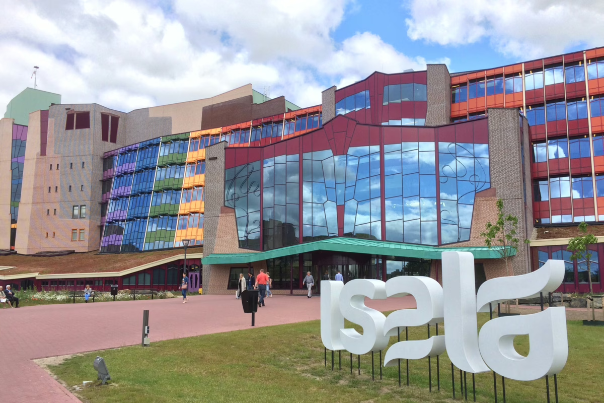
Health economists predict that the number of patients in Western Europe will double in the next 20 to 25 years. This will of course affect radiology, which will have to prepare for a corresponding increase in the number of examinations and findings. “If the predictions come true, we will have to become more efficient in what we do – as I do not expect the same growth in the number of radiologists. I see artificial intelligence (AI) as part of the possible solution. This applies not only to radiology, but to all activities along the care process,” says Dr. Martijn F. Boomsma, Radiologist at Isala in Zwolle. The Isala hospital group also operates smaller facilities in Meppel, Steenwijk, Kampen and Heerde. The group has a total of 1,200 beds and is one of the largest non-academic teaching hospitals in the Netherlands. The department of Medical Imaging at Isala examines over 1,200 radiological examinations per day.

The Dutch Radiological Society has also defined AI as one of the four most important developments for the next decade, and indeed radiology is already a pioneer in the application of this technology. And that’s a good thing, says Dr. Boomsma: “AI, in combination with an experienced radiologist, increases diagnostic accuracy because it can effectively support and relieve radiologists in certain aspects.” However, clinics and manufacturers would have to prove that the algorithms have a concrete positive impact on health outcomes. This could greatly improve clinical adoption as it would make a clear case for reimbursement for AI and getting AI into national guidelines. Dr. Boomsma believes his discipline is on the right track, even if it still faces a number of hurdles.

Committed partnership at eye level

The department of Medical Imaging has been using diagnostic AI applications for four years and has been using contextflow ADVANCE Chest CT since the end of 2022. Isala runs the solution on its own servers and has integrated it with its image data management system (PACS) from Sectra.

Implementing the AI algorithms was not a plug-and-play process. The hurdle was not only the vendors, but also internal processes. “We had to convince several parties and get the go-ahead from many echelons before we could start. It took a year before the application was really ready for operation, fully integrated, reliable, and with an uptime of 97 percent. The technical integration was then very simple and straightforward,” says Dr. Boomsma. This process requires a lot of commitment and perseverance from everyone involved. But it is also the point at which trust is built. “This is where true partnership shows itself, and we are still experiencing this with contextflow,” the radiologist is pleased to say. 

Dr. Boomsma also notes contextflow’s effort to continuously develop its solution in close cooperation with its users in order to optimize the workflow and thus increase diagnostic value. “This was also a decisive reason for us to choose contextflow. We see a high level of professionalism and agility, as well as the company’s vision to get the best out of AI in thoracic imaging,” he reports. This is also evident in their day-to-day interactions. “Employees respond promptly to inquiries and problems by phone or email. To this end, they always think in terms of solutions and work to solve problems as quickly as possible,” the radiologist elaborates. ADVANCE Chest CT itself stands out because it can be operated without much training. “With a little practice, you can use the software very quickly; it’s user-friendly. But it also changes the way you look at things,” Dr. Boomsma says.

Valuable support for the findings

Dr. Boomsma has developed his own approach to using the AI software. First, he looks at the key images and obtains a global overview. Then he reads the scan, incorporating the referring physician’s specific questions into the findings and preparing his report. “ADVANCE Chest CT helps me identify and quantify nodules. This allows me to clearly identify whether the disease is progressive or stable. Of course, I can also discard or change individual results I don’t consider relevant, as they are false positives or do not relate to the specific question,” says Dr. Boomsma, explaining his workflow. Not only does he use the AI for pulmonary nodules, but also for the detection and quantification of emphysema.

Meanwhile, ADVANCE Chest CT is an important support for reporting at Isala. “On an CT image without contrast, it is very difficult to find a four-millimeter lesion in the perihilar region. But the system reliably shows it to me. It allows me to make sure I haven’t missed anything. It also helps us in our daily work. For example, it is reassuring to know that the AI is always by your side during a nighttime emergency scan and to be sure that everything important can be reported,” says Dr. Boomsma. He also sees the potential to speed up the reporting of findings, citing scans without significant findings as an example. The algorithm can mark these as such, so that the radiologist only has to check them once and can then concentrate on more complex cases.

More than other AI algorithms

“ADVANCE Chest CT gives me much more information than AI solutions from other vendors. It detects and quantifies nodules, emphysema and also fibrosis. The precise information on the extent of manifestation of suspected pathology adds real value to the findings because it can be indicative and correlate with the patient’s condition and  may guide the need for therapy,” emphasizes Dr. Boomsma. Consistency of findings in general remains a challenge, due to different scan settings, inspiration etc. He highlights ADVANCE Chest CT’s SEARCH feature as another unique selling point. With a single mouse click, an overview of similar cases opens from an extensive database, which radiologists can use to support their diagnosis.

The TIMELINE module is a further facilitator. It clearly visualises the changes in the detected nodules over time showing the percentage of growth as well as the time of volume doubling. This is particularly helpful when preparing for tumour boards and multidisciplinary meetings. The integration with the Sectra PACS gives the potential to seamlessly incorporate the results into the report.

Other News

AI Interest Group for Imaging (AIGI) Task Force
2023-08-07

The “Interest Group for AI in Imaging” (AIGI) started its work in January and advocates the optimization and greater use of IHE profiles when using AI (Artificial Intelligence). 

AI applications are slowly becoming the standard in radiological reporting, but there’s a lack of standardization when it comes to the implementation of AI. Two IHE (Integrating the Healthcare Enterprise) integration profiles already exist (IHE AIW-I & IHE AIR) for the integration of artificial intelligence (AI) and its communication with DICOM data, but so far there are only very few implementations in current products visible. Thus, Marc Kämmerer, member of the IHE Europe Steering Committee and Head of Innovation Management at VISUS Health IT GmbH, initiated AIGI.

What is AIGI?

AIGI is a task force of IHE Europe consisting of radiology AI users, software and PACS vendors, marketplace operators as well as other interest groups. Its main interest and goal is to define the means for a standardized and applicable data workflow for actual use cases in European healthcare systems, including how to:

  • deploy and maintain AI applications
  • connect AI applications with end users’ systems
  • integrate the AI application output in end-users’ systems
  • collect and provide end user feedback

Examining the entire workflow between user and AI

The demand for standardization is high from all sides of the workflow equation. AIGI is examining the entire process chain between the user and the AI solution for practical applicability and feasibility on the basis of the profiles mentioned. Looking for holes in the existing standards, the group will create proposals and frameworks for improved, bi-directional data flow between AI and the user.

One initial area of focus: currently when a radiologist sends DICOM data to an AI software, there is no feedback as to how long it will take for the result to reach the PACS. Should the radiologist wait or start with the next patient? A status query could be integrated as standard via the IHE AIW-I profile, and that is exactly the type of question AIGI hopes to standardize.

The same applies to error messages, which previously only took the form of empty reports or system crashes. Similar to DICOM email, feedback should be given here as to whether and what type of error has occurred. In terms of interoperability, the content of these messages would have to be defined so that they have the same meaningfulness independent of the display system. 

Another example: When reporting, it can make sense to play certain evaluations prominently on the surface – for example, degrees of malignancy in mammography. In principle, this is possible via DIOCM-SR objects, but here too the challenges lie in the details.

Simplified AI access for all

“Working on these standards is very important in order to implement binding structures in the AI ​​processes as early as possible. We are currently noticing that many AI providers and AI marketplace operators are working with APIs. However, it is impossible for PACS manufacturers to use all the APIs available on the market. There are currently well over 20 AI marketplace operators and several hundred AI providers. Here we have to find solutions quickly that ultimately benefit everyone involved – manufacturers and users. From the positive response to our task force, we can see that fortunately all parties involved see things the same way. And it didn’t take long for the IHE to convince the group to set up the group,” says Marc Kämmerer, pleased with the response from industry and practice.

contextflow is a proud supporter of AIGI. Currently we are participating in the subgroup related to Longitudinal Data, Reporting, and Pseudo-/Anonymization. The AIGI Taskforce is open to additional members. Its intended output are best practice white papers, correction proposals, and work item proposals to ultimately benefit users, PACS and AI manufacturers and AI marketplace operators. For more information, click here.

Other News

contextflow approved for EIC Accelerator Equity Investment
2023-06-22

We are beyond thrilled to announce that contextflow’s application for the European Innovation Council (EIC) Accelerator equity investment program has been accepted!!!

The latest round of EIC funding saw stiff competition: 139 companies were interviewed by juries of experienced investors and entrepreneurs, out of a total of 551 full proposals submitted. We are the only Austrian company selected in the latest round of funding.

contextflow’s history with the EIC dates back to 2020, when we became the fortunate recipients of a €1.2 Million EIC grant. This shows a clear commitment from the European Commission to support European-born companies with cutting-edge innovation over the long-term. The trust bestowed upon us does not go unnoticed; the best way to show gratitude is to continue to develop our comprehensive clinical decision support for chest CT to enable radiologists to perform their routine tasks faster and with higher certainty and accuracy.

An additional HUGE thanks our early supporters and stakeholders for taking us from university spinoff to the 40+ member team we are today.

Technische Universität Wien
Medizinische Universität Wien
TU Wien Innovation Incubation Center (i²c)
INiTS | Vienna’s High-Tech Incubator
Health Hub Vienna
LISAvienna – Life Science Austria Vienna
FFG Österreichische Forschungsförderungsgesellschaft mbH
Vienna Business Agency
Austria Wirtschaftsservice

Special personal thanks to Angelo Nuzzo, PhD, MBA, MA, Birgit Hofreiter, Alexandra Negoescu & Irene Fialka for guiding us from the very beginning. We couldn’t have done it without your support!

Other News

Radiology Symposium Mainz returns for a 2nd year
2023-08-09

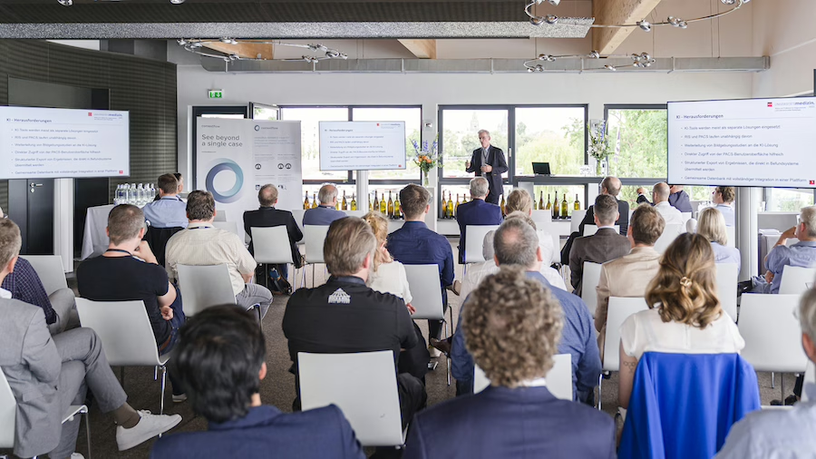
An impressive crowd at this year’s Radiology Symposium Mainz! For a second year running, we gathered at the Bootshaus Mainz at the end of June to discuss current radiology AI topics.

Thank you to our fabulous radiology speakers & partners, starting with host Prof. Dr. med. Peter Mildenberger from University Medical Center Mainz. Univ.-Prof. Dr. med. Christoph DĂĽber, Prof. Dr. med. Mike Notohamiprodjo, Elodie Weber, Erwin Krikken, Karin Klein, Marcel Wassink, Mark Rawanschad, PD Dr. Christian Elsner, Florian Brandt, Knut Dietrich-Thiel, Bernd SchĂĽtze, Alex Lemm, Barbara Lampl, PD Dr. Daniel Pinto dos Santos, Prof. Dr. med. Elmar Kotter, medavis GmbH, EIZO Healthcare, DFC-SYSTEMS GmbH.

We would also like to thank our friends at Sectra for their continued collaboration!

Hope to see you again next year!

Other News

Save time and improve diagnostic quality
2023-05-17

contextflow ADVANCE Chest CT improves lung diagnostics at St. Bernhard-Hospital Kamp-Lintfort

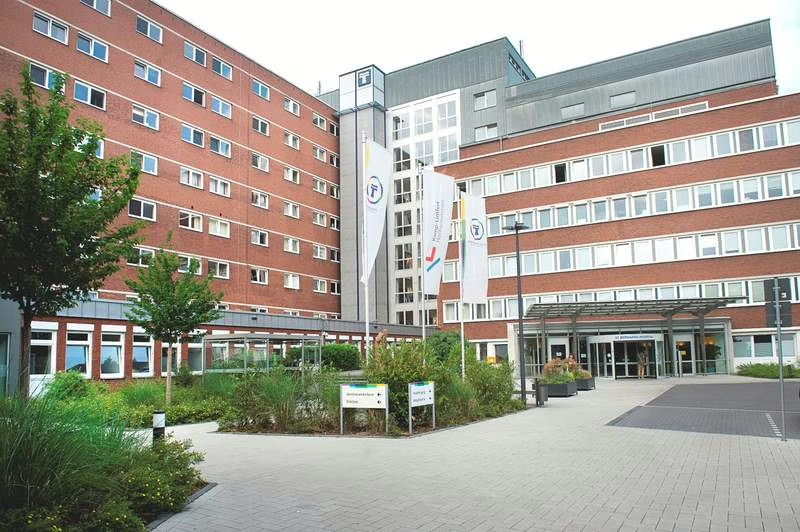
PD Dr. Hilmar KĂĽhl was employed at the University Medical Center Essen for 16 years, half of this time at the Ruhrlandklinik, the major West German lung center. He now brings this expertise to bear as head physician of the Department of Radiology at St. Bernhard-Hospital Kamp-Lintfort. The regional provider with 356 beds treats around 15,000 inpatients and 30,000 outpatients annually. 

As a recognized thoracic radiologist, Dr. KĂĽhl draws his patients to Kamp-Lintfort from a fairly large area between Wesel, Neuss, Duisburg and Straelen. The requirements range from outpatient questions and the primary diagnosis of various lung diseases to the staging of bronchial carcinoma. Together with his team, he performs up to 1,500 chest CTs per year.

In 2015 while still at the University Medical Center Essen, the chief radiologist had his first contact with artificial intelligence (AI) methods in thoracic diagnostics. Since June 2022, he has been working with ADVANCE Chest CT, contextflow’s AI solution for detecting parenchymal changes in the lungs. “We are a member of the West German Teleradiology Network, which offers the solution via its AI marketplace. Via our image data management system (PACS) JiveX from VISUS, we can then use the algorithm on a pay-per-use basis,” Dr. KĂĽhl explains the construct. Advantages: no software installation, guaranteed data protection and secure communication infrastructure.

Automated integration into the workflow

The radiologists at St. Bernhard Hospital have defined examinations that are always subject to analysis by the AI. As soon as the questions “chronic bronchitis”, “COPD”, “pulmonary skeleton changes” and “fibrosis” appear, the image data is automatically sent to the platform of the West German teleradiology network and analyzed with ADVANCE Chest CT. The result is also automatically fed back into the PACS. “These automations are extremely helpful because they save us time-consuming, manual activities,” says Dr. KĂĽhl, citing one advantage of the process. “If our radiologists were to post-process and analyze the images, this would take up to ten minutes per examination. The AI analyzes a total of 19 image patterns in significantly less time and also provides me with differential diagnoses.” In addition, it is also possible to display reference images.

The chief radiologist particularly appreciates the integration of the algorithm into the workflow. “Every mouse click means extra work and costs time. We save that with the solution we use,” Dr. KĂĽhl emphasizes. He is also impressed by the feedback, where all analysis results are clearly displayed on a PDF page. “This allows me to identify the relevant information very quickly and include it in the findings. The differential diagnoses are also very helpful.”

Added value for daily work

Lung parenchymal diseases, especially COPD with emphysema and interstitial parenchymal diseases, play a major role in diagnostics at St. Bernhard-Hospital Kamp-Lintfort. ADVANCE Chest CT detects and quantifies each of these pathologies. In selected patients, Dr. Kuehl applies a computer-assisted diagnosis (CAD) tool in addition to AI for comparison purposes. Using the CAD system does present challenges, however, as the chief radiologist elaborates: “That’s when we use two modules: One provides information on the extent of emphysema, the other identifies pulmonary nodules. In total, a radiologist is quickly occupied for 15 minutes. ADVANCE Chest CT delivers both results together – and even more, for example, information on infiltrates. This means an immense reduction in workload and time savings for us.”

In principle, AI solutions always raise the question of the data on which the learning process is based and how close they are to the ground truth, i.e. the verified clinical ground truth. This is particularly complicated as it relates to the differentiation of changes in the lung, where image impression and clinical relevance do not always coincide. On CT, various parenchymal patterns besides emphysema regularly come into play here, such as ground-glass opacities and honeycombing, as well as interstitial changes such as traction bronchiectasis or reticular patterns. “Quantification in particular supports the diagnosis and increases the certainty of the findings,” emphasizes Dr. KĂĽhl.

Playing to the strengths of AI

In general, ADVANCE Chest CT helps him to produce a very high quality report in less time. The gain in speed primarily comes from quantifying the pathological changes, which must be done manually without IT support. Detection and quantification are crucial for therapy, which ranges from the application of a spray to surgery. “And if I can then provide the attending physician with reliable data, this increases the value of my findings in a relevant way and improves the therapy for the patient. Last but not least, this leads to increased satisfaction among clinicians and referring physicians,” says Dr. KĂĽhl.

Radiologists also expect real added value when diagnosing rare diseases. Here the AI can support less experienced colleagues by pointing out possible differential diagnoses based on analyzed parameters – as ADVANCE Chest CT already does today. “This simplifies the path from pattern quantification to diagnosis. I’m offered a reference pattern and told in what percentage of cases it is verified with a specific disease. On the one hand, limiting the differential diagnoses simplifies the reporting, but on the other hand, it also raises the quality of the findings in a relevant way. This can make it possible to bring AI to a wider audience.” The work is facilitated by the fact that the software is self-explanatory and easy to use even after a brief introduction.

In the context of the close cooperation, PD Dr. Hilmar KĂĽhl perceives contextflow as an extremely committed partner. “The company has a genuine interest in our feedback and values its clinical partners who use the software in their daily routine. Accordingly, contextflow has also supported me very well throughout the process.” It sounds like the collaboration has a bright future.

Other News

Befundungszeit sparen und Befundqualität heben
2023-06-07

ADVANCE Chest CT von contextflow verbessert Lungendiagnostik im St. Bernhard-Hospital Kamp-Lintfort

PD Dr. Hilmar KĂĽhl war 16 Jahre lang an der Universitätsmedizin Essen beschäftigt, die Hälfte dieser Zeit an der Ruhrlandklinik, dem groĂźen westdeutschen Lungenzentrum. Diese Expertise bringt er nun auch als Chefarzt der Klinik fĂĽr Radiologie im St. Bernhard-Hospital Kamp-Lintfort ein. Der regionale Versorger mit 356 Betten versorgt jährlich rund 15.000 Patienten stationär und 30.000 ambulant. 

Als anerkannter Thoraxradiologe zieht Dr. Kühl seine Patienten aus einem recht großen Gebiet zwischen Wesel, Neuss, Duisburg und Straelen nach Kamp-Lintfort. Die Anforderungen reichen von ambulanten Fragestellungen und die Primärdiagnostik diverser Lungenerkrankungen bis zum Staging des Bronchialkarzinoms. Mit seinem Team befundet er bis zu 1.500 Thorax-CTs pro Jahr.

2015 hatte der Chefradiologe erstmals Berührung mit Verfahren der Künstlichen Intelligenz (KI) in der Thoraxdiagnostik, damals noch an der Universitätsmedizin Essen. Seit Juni 2022 arbeitet er nun mit ADVANCE Chest CT, der KI-Lösung von contextflow zur Detektion von Parenchymveränderungen der Lunge. „Wir sind Mitglied im Westdeutschen Teleradiologieverbund, der diese Lösung über seinen KI-Marktplatz anbietet Über unser Bilddatenmanagementsystem (PACS) JiveX von VISUS können wir den Algorithmus dann im Pay-per-Use-Verfahren nutzen“, erläutert Dr. Kühl das Konstrukt. Vorteile: keine Softwareinstallation, gewährleisteter Datenschutz und sichere Kommunikationsinfrastruktur.

Automatisiert in den Workflow integriert

Die Radiologen im St. Bernhard-Hospital haben CT-Untersuchungen definiert, die immer einer Analyse durch die KI unterzogen werden. Sobald die Fragestellungen „chronische Bronchitis“, „COPD“, „Lungengerüstveränderungen“ und „Fibrose“ auftauchen, werden die CT-Bilddaten automatisch an die Plattform des Westdeutschen Teleradiologieverbundes geschickt und mit ADVANCE Chest CT analysiert. Das Ergebnis wird ebenfalls automatisch in das PACS zurückgespielt. „Diese Automatismen sind extrem hilfreich, weil sie uns zeitraubende manuelle Tätigkeiten ersparen“, nennt Dr. Kühl einen Vorteil des Verfahrens. „Würden unsere Radiologen die Bilder nachbearbeiten und analysieren, würde das pro Untersuchung bis zu zehn Minuten dauern. Die KI analysiert in deutlich geringerer Zeit insgesamt 19 Bildmuster und liefert mir zudem Differenzialdiagnosen.“ Darüber hinaus ist es möglich, sich auch Referenzbilder anzeigen zu lassen.

Der Chefradiologe schätzt besonders die Integration des Algorithmus in den Workflow. „Jeder Mausklick bedeutet Mehrarbeit und kostet Zeit. Die ersparen wir uns mit der genutzten Lösung“, betont Dr. Kühl. Auch die Rückmeldung, bei der auf einer PDF-Seite alle Analyseergebnisse übersichtlich dargestellt sind, überzeugt ihn. „So kann ich sehr schnell die relevanten Informationen identifizieren und in den Befund aufnehmen. Sehr hilfreich sind auch die Differenzialdiagnosen.“

Mehrwerte für die tägliche Arbeit

Bei der Diagnostik im St. Bernhard-Hospital Kamp-Lintfort spielen die Lungenparenchymerkrankungen, insbesondere die COPD mit Emphysem und interstitielle Parenchymerkrankungen eine große Rolle. ADVANCE Chest CT detektiert und quantifiziert jede dieser Pathologien. Bei ausgewählten Patienten wendet Dr. Kühl neben der KI auch ein Tool zur computerassistierten Diagnose (CAD) zu Vergleichszwecken an. Die Verwendung des CAD-Systems bringt allerdings Herausforderungen mit sich, wie der Chefradiologe ausführt: „Da setzen wir dann zwei Module ein: Das eine liefert Aussagen zur Ausprägung des Lungenemphysems, das andere identifiziert Lungenrundherde. Insgesamt ist da ein Radiologe schnell 15 Minuten beschäftigt. ADVANCE Chest CT liefert beide Ergebnisse zusammen – und noch mehr, beispielsweise Angaben zu Infiltraten. Das bedeutet für uns eine immense Arbeitserleichterung und Zeitersparnis.“

Grundsätzlich stellt sich bei KI-Lösungen immer die Frage, auf welcher Datengrundlage der Lernprozess erfolgt bzw. wie dicht sie dabei an der sogenannten Ground Truth, also der verifizierten klinischen Grundwahrheit, sind. Gerade bei der Differenzierung von Veränderungen am Lungengerüst, wo sich Bildeindruck und klinische Relevanz nicht immer decken bzw. ähnliche klinische Symptome sehr verschiedene CT-Morphologie aufweisen können, ist das kompliziert. Im CT kommen hier regelhaft verschiedene Parenchymmuster neben dem Emphysem ins Spiel; wie etwa Milchglas-Infiltrate, Wabenbildungen sowie interstitielle Veränderungen wie Traktionsbronchiektasen oder retikuläre Muster. „Das löst contextflow mit seiner Lösung allerdings sehr gut. Gerade die Quantifizierung unterstützt die Diagnosestellung und erhöht die Befundsicherheit“, betont Dr. Kühl.

Stärken der KI ausspielen

Ganz allgemein hilft ihm ADVANCE Chest CT dabei, in kürzerer Zeit einen Befund von sehr hoher Qualität zu erstellen. Der Geschwindigkeitsgewinn ergibt sich primär bei der Quantifizierung der krankhaften Veränderungen, die ohne IT-Unterstützung manuell vorgenommen werden müssen. Detektion und Quantifizierung sind entscheidend für die Therapie, die von der Anwendung eines Sprays bis zur Operation reicht. „Und wenn ich dem behandelnden Arzt dann verlässliche Daten an die Hand geben kann, steigert das den Wert meines Befundes auf relevante Weise und verbessert die Therapie für den Patienten. Das führt nicht zuletzt zu einer steigenden Zufriedenheit der Kliniker und Zuweiser“, so Dr. Kühl.

Einen echten Mehrwert verspricht sich der Radiologe auch bei der Diagnostik seltener Erkrankungen. Da kann die KI besonders weniger erfahrene Kollegen unterstützen, indem der Algorithmus aufgrund analysierter Parameter auf mögliche Differenzialdiagnosen hinweist – so wie ADVANCE Chest CT es heute bereits tut. „Das vereinfacht den Weg von der Musterquantifizierung zur Diagnose. Mir wird ein Referenzmuster angeboten und gesagt, in wie viel Prozent der Fälle es mit einer spezifischen Erkrankung verifiziert ist. Die Einschränkung der Differenzialdiagnosen vereinfacht einerseits die Befundung, hebt andererseits aber auch die Befundqualität relevant an. Damit kann es gelingen, die KI in die Breite zu tragen.“ Erleichtert wird die Arbeit dadurch, dass die Software selbsterklärend und bereits nach einer kurzen Einführung leicht zu bedienen ist.

Im Rahmen der engen Zusammenarbeit nimmt PD Dr. Hilmar Kühl contextflow als äußerst engagierten Partner wahr. „Das Unternehmen hat ein wirkliches Interesse an unserem Feedback und schätzt seine klinischen Partner, die die Software in ihrem Routinealltag nutzen. Dementsprechend hat contextflow mich auch im gesamten Prozess sehr gut unterstützt.“ Das klingt danach, dass die Zusammenarbeit eine gute Zukunft hat.

Other News

contextflow in clinical use showcased at ECR 2023 in Vienna
2023-07-20

contextflow was featured in a few scientific presentations at the European Congress of Radiology (ECR) 2023 in Vienna showcasing the clinical efficacy of their ADVANCE Chest CT AI solution in lung disease diagnosis and its valuable insights for clinical decision-making.

One of the key research featured in the scientific presentation at the conference explored the relationship between lung volumetrics obtained from contextflow’s AI solution and lung function tests correlating with fibrosis progression. The combination of lung quantification values of reticulation and honeycombing, along with blood monocyte count, has been identified as a potential biomarker for predicting the progression of lung fibrosis within 12 months.

In another one, contextflow’s AI solution was shown to be effective in quantifying disease patterns in lung CT scans associated with individual mortality outcomes in idiopathic pulmonary fibrosis. The study revealed that lung volumetrics obtained from the AI solution were able to predict the outcomes of fibrotic patients. Both research showed the potential of contextflow’s AI solution as a valuable tool for predicting disease progression in patients with fibrotic lung diseases.

contextflow presented research comparing AI measures of emphysema with Hounsfield Unit (HU) emphysema measures. The study demonstrated that the AI solution provided accurate and reliable measures of emphysema, offering a promising alternative to traditional HU-based measurements.

Moreover, contextflow’s AI solution was found to be a valuable tool in predicting complications after CT-guided needle biopsy in the lungs, as presented by the team from Mainz in a study titled “Pre-interventional AI-supported Automated Lung Parenchyma Quantification Predicts Post-interventional Complications in CT-guided Lung Biopsies”. The study showed that lung volumetrics obtained from the AI solution could help identify patients at risk of post-interventional complications, allowing for better patient management and improved outcomes.

Finally, the team from Jönköping delivered a poster presentation on the use of contextflow’s ADVANCE Chest CT in clinical settings.The findings from the three-month follow-up indicate that the AI solution has the potential to significantly reduce reading times, with a reduction of 17% and 26% observed for emergency/in-house and elective exams respectively. The solution was in use for a relatively short time at the hospital, but it indicated the potential of the AI solution to streamline workflow and improve efficiency in clinical practice.

Other News

contextflow to bolster ADVANCE Chest CT with incidental pulmonary embolism (IPE)
2023-02-27

The inclusion of IPE will help strengthen the company’s offerings as a market leader in comprehensive computer-aided detection support for chest CT

28.2.23 Vienna, Austria – The Vienna-based chest experts at contextflow are announcing a new feature at this year’s European Congress of Radiology: incidental pulmonary embolism detection. According to Chief Product Officer Markus Krenn, “Radiologists requested the IPE feature because of the critical nature of pulmonary embolism. By fulfilling their request, we continue to build trust and help patients in the process.”

contextflow offers comprehensive computer-aided detection software for chest CT to support the diagnosis and treatment monitoring of lung cancer, ILD and COPD. IPE will be added to the company’s core product, ADVANCE Chest CT, which automatically detects, quantifies and visualizes 8 disease patterns including lung nodules, displaying relevant information directly in the radiologist’s PACS viewer. In addition, contextflow’s TIMELINE feature quickly and objectively tracks changes in lung nodules over time, currently a very difficult and time-consuming task for radiologists.

Incidental pulmonary embolism occurs when a patient is being scanned for reasons other than PE. Thus, there is a risk to the patient if there is any delay in reporting. The rates of missed IPE are relatively high, and it appears that more and more radiology departments are looking into tools to reduce these figures.

With this new feature, contextflow aims to increase the speed of care for patients with IPE and ensure radiologists get the comprehensive support for the assessment of chest CTs they need. Again Chief Product Officer Markus Krenn, “We work very closely with a group of practicing radiologists who provide us with a constant feedback loop. We take their requests very seriously, and will continue to add new features in the future accordingly.”

Other News

Radiologist Interview Series – Prof. Dr. Peter Mildenberger
2023-02-17

The PACS as an Integration Platform for AI Solutions

Interview with Prof. Dr. Peter Mildenberger, Senior Physician and IT Officer at the Department of Radiology at Mainz University Medical Center.

Professor Mildenberger, where do you see the potential of artificial intelligence in radiology?

Prof. Dr. Peter Mildenberger: In principle, AI tools already offer many opportunities to improve the quality of diagnostics as well as to allow quantifications that we do not typically make in the data. I can imagine AI systems, for example, in the diagnosis of pulmonary nodules or liver metastases, for the determination of organ volumes or in the analysis of body composition.

How far has AI come today?

Prof. Dr. P. Mildenberger: I do not dare to make a final assessment. We have had experience with some tools, but I don’t have a complete overview. There are a lot of systems, and for me the exciting question is which of them will find their way into clinical routine and hold their own there.

What does AI have to offer in order to be accepted in clinical routine?

Prof. Dr. P. Mildenberger: There are various aspects. In the case of pulmonary nodule detection, for example, the practical issue is the number of false-positive findings. Then acceptance certainly stands or falls with seamless integration into the workflow. contextflow enables the automated import of results into the PACS – not as images, but as values. Only when the radiologist has validated this and created his report, however, does the referring physician have access to it. I find it problematic to leave clinicians alone with the results of AI.

How can AI algorithms be brought into the clinics?

Prof. Dr. P. Mildenberger: There are different models. In the end, it depends on how often individual tools are used. Is it worth buying a software solution or should the pay-per-use model be preferred? The latter is certainly attractive if I want to use different algorithms, but not so frequently. The IT infrastructure of the company also plays a decisive role. Here in Mainz, we operate the IT for radiology ourselves and consequently have few problems integrating new software solutions. This is usually quite different in a medium-sized or smaller hospital. It can then make sense to connect a platform and use it to access various algorithms. Before that, however, each institution must clarify whether the use of cloud solutions is an option – something we have rejected in Mainz so far.

What is your experience with AI applications?

Prof. Dr. P. Mildenberger: It is too early to make a general assessment. We have only tried out a fairly small spectrum of applications so far. We lack experience with essential tools, such as for detecting fractures or pulmonary embolisms. We certainly have the most comprehensive experience with pulmonary nodules. We don’t have this fixed in the workflow, but when a colleague has looked at his lung CT, the AI takes a second look at each image. That’s quite interesting and sometimes helpful.

What is your AI strategy?

Prof. Dr. P. Mildenberger: We rely on Sectra’s image data management and use the PACS as a facility-wide platform. Specifically for CT and MRI image processing, we use syngo.via from Siemens Healthineers. Any AI tools we acquire must integrate with these platforms, i.e., accept information and transmit results back. 

Right now, we are looking at identifying modules that we want to use in the future. For the neuroradiologists, these could be systems for MS diseases; we radiologists primarily see support for pulmonary embolisms as well as prostate and breast MRI. We will look at appropriate modules for the respective clinical applications, then decide individually and buy the solution. I still don’t see a platform solution for us.

Thank you very much for the informative discussion, Professor Mildenberger.

Other News