
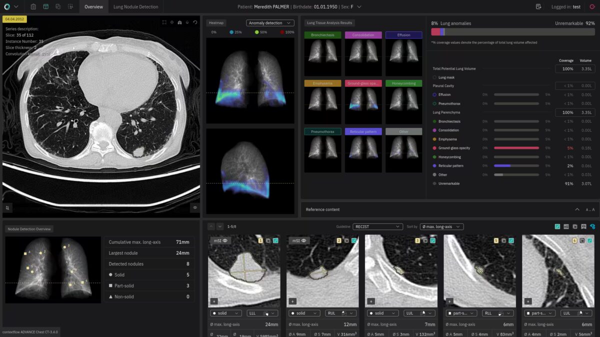
Evaluating chest CTs can be a complex and time-consuming process. So we’ve packed contextflow ADVANCE Chest CT with qualitative and quantitative insights to help you objectively report on suspected lung cancer, ILD, and COPD cases. Over time. From within your native viewer.
DETECT / Nodule Detection

Features
- Detection & quantification of nodules (4-30mm diameter) from within your native viewer.
- Nodule classification (solid, part-solid, non-solid)
- We detect and measure nodules in accordance with the following guidelines:
- British Thoracic Society guidelines for the investigation and management of pulmonary nodules
- European Union Position Statement on Lung Cancer Screening
- Guidelines for Management of Incidental Pulmonary Nodules Detected on CT Images: From the Fleischner Society 2017
- International Early Lung Cancer Action Program: Screening Protocol, 2021
- Lung-RADS Version 1.1 2019
- Response Evaluation Criteria in Solid Tumors (RECIST)
- For USA only: the DETECT feature is currently under development as a standalone product – not yet cleared for clinical use.
TIMELINE / Nodule Tracking

Features
- Tracks changes in detected nodules over time, regardless of the number of priors
- Indicates growth percentage and volume doubling time
- Helps to prepare for tumor boards and multidisciplinary board meetings
- TIMELINE is currently only available for nodule tracking. Tracking of disease patterns over time is coming in 2023.
INSIGHTS / Lung Tissue Analysis

Features
- Anomaly heatmaps indicating overall distribution of disease patterns
- Quantification of total lung volume affected by disease patterns
- Quantification & individual heatmaps for 7 key image findings:
- Consolidation
- Effusion
- Emphysema
- Ground-glass opacity
- Honeycombing
- Pneumothorax
- Reticular Pattern
iCAC *Coming Soon*

Features
- Total volume of detected coronary artery calcifications
- Incidental cardiovascular disease risk categorisation
- Estimated Agatston score
SEARCH / 3D Image Search

Features
- Detection & analysis of 19 image patterns
- Airway wall thickening
- Atelectasis
- Bronchiectasis
- Bulla
- Consolidation
- Cyst
- Effusion
- Emphysema
- Ground-glass opacity
- Honeycombing
- Mass
- Mosaic attenuation pattern
- Nodular pattern
- Nodule
- Pneumothorax
- Pulmonary cavity
- Reticular pattern
- Tree-in-bud
- Unremarkable: includes patterns with no evidence of pathological changes and currently not explicitly incorporated
- Links to differential diagnosis literature
- Retrieval of similar cases to yours from a curated knowledge base
- Validated average report reading time savings 31%
Testimonials
I have been following contextflow’s progress practically since the company’s founding, and their traction in the area of chest CT is impressive. Being able to shape clinical decision support tools that myself and colleagues can benefit from in clinical practice is a big motivator. We’re literally shaping the future.
Jacob Visser
Chief Medical Information Officer & Head of Imaging IT and Value-Based Imaging at Erasmus MC

I really like the transparency of contextflow as opposed to other black box AI solutions. It’s designed to support my workflow while leaving the final decision up to me.
Elmar Kotter
Vice Chair and Head of Imaging Informatics at the Department of Radiology at Freiburg University Medical Center
In the world of AI, it’s crucial to use it safely, be clear about what it does, and make ethical choices. This means moving forward with innovation in a responsible way, creating a future that’s both advanced and thoughtful.
Geraldine Dean
MD MSc MRCS FRCR Consultant Radiologist, Artificial Intelligence Lead NHS SW London Imaging Network
For interstitial lung disease, we use contextflow on a daily clinical basis. We now put the major information into the radiology report. And this is what our clinicians expect from us: to be able to quantify the disease and especially to quantify disease progression in order to improve clinical decision making.
Gerlig Widmann
Managing Senior Physician at the University Department of Radiology at the Medical University of Innsbruck

One of the great features of contextflow is the TIMELINE view, which offers the possibility to actually analyse follow-up scans. And that has a lot of value for our clinical practice because patients will return to our practice for follow-up imaging.
Willem Grootjans
Head of the Imaging Services Group at the Department of Radiology, Leiden University Medical Center
I use contextflow in any routine scan performed, for example, for staging, or for other disease evaluation. It helps me a lot to recognize patterns in patients where you’d not expect or where we cannot clearly see the pathology behind it. So it helps us a lot as a double checker.
Lukas Müller
Radiology Resident & Clinician Scientist at the Medical University of Mainz
contextflow is one of the applications that certainly fits radiology’s current needs and can simplify the analysis of complex lung pathology. With the right insights and technology, we can succeed in introducing AI in a very attractive way to radiology departments on a global scale.
Erik Ranschaert
Former President of the European Society of Medical Imaging Informatics (EuSoMII), Radiologist at St. Nikolaus Hospital in Eupen
We’re very interested in using AI to improve the hospital experience for both doctors and patients; contextflow’s use of deep learning, particularly for lung diseases, is exactly the type of technology we want to evaluate. I very much look forward to the results.
Christian Herold
Head of Radiology at Vienna General Hospital
After using and advising several radiology AI software companies, I can say that what contextflow offers is actually the next generation of AI products to support the radiologist, not replace them. Their general approach means they recognize all relevant findings, not just one.
Anand Patel
MD, Chief of Interventional Radiology, Providence Little Company of Mary Medical Centers
We are very interested in using tools based on artificial intelligence like contextflow to support the decision in the diagnostic process based on the image.
Lluís Donoso Bach
President of the International Society of Radiology