AI Interest Group for Imaging (AIGI) Task Force

The « Interest Group for AI in Imaging » (AIGI) started its work in January and advocates the optimization and greater use of IHE profiles when using AI (Artificial Intelligence). 

AI applications are slowly becoming the standard in radiological reporting, but there’s a lack of standardization when it comes to the implementation of AI. Two IHE (Integrating the Healthcare Enterprise) integration profiles already exist (IHE AIW-I & IHE AIR) for the integration of artificial intelligence (AI) and its communication with DICOM data, but so far there are only very few implementations in current products visible. Thus, Marc Kämmerer, member of the IHE Europe Steering Committee and Head of Innovation Management at VISUS Health IT GmbH, initiated AIGI.

What is AIGI?

AIGI is a task force of IHE Europe consisting of radiology AI users, software and PACS vendors, marketplace operators as well as other interest groups. Its main interest and goal is to define the means for a standardized and applicable data workflow for actual use cases in European healthcare systems, including how to:

  • deploy and maintain AI applications
  • connect AI applications with end users’ systems
  • integrate the AI application output in end-users’ systems
  • collect and provide end user feedback

Examining the entire workflow between user and AI

The demand for standardization is high from all sides of the workflow equation. AIGI is examining the entire process chain between the user and the AI solution for practical applicability and feasibility on the basis of the profiles mentioned. Looking for holes in the existing standards, the group will create proposals and frameworks for improved, bi-directional data flow between AI and the user.

One initial area of focus: currently when a radiologist sends DICOM data to an AI software, there is no feedback as to how long it will take for the result to reach the PACS. Should the radiologist wait or start with the next patient? A status query could be integrated as standard via the IHE AIW-I profile, and that is exactly the type of question AIGI hopes to standardize.

The same applies to error messages, which previously only took the form of empty reports or system crashes. Similar to DICOM email, feedback should be given here as to whether and what type of error has occurred. In terms of interoperability, the content of these messages would have to be defined so that they have the same meaningfulness independent of the display system. 

Another example: When reporting, it can make sense to play certain evaluations prominently on the surface – for example, degrees of malignancy in mammography. In principle, this is possible via DIOCM-SR objects, but here too the challenges lie in the details.

Simplified AI access for all

“Working on these standards is very important in order to implement binding structures in the AI ​​processes as early as possible. We are currently noticing that many AI providers and AI marketplace operators are working with APIs. However, it is impossible for PACS manufacturers to use all the APIs available on the market. There are currently well over 20 AI marketplace operators and several hundred AI providers. Here we have to find solutions quickly that ultimately benefit everyone involved – manufacturers and users. From the positive response to our task force, we can see that fortunately all parties involved see things the same way. And it didn’t take long for the IHE to convince the group to set up the group, » says Marc Kämmerer, pleased with the response from industry and practice.

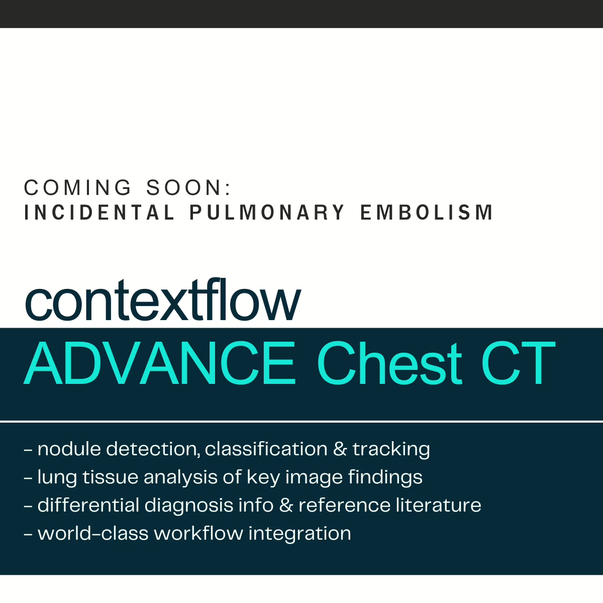
contextflow is a proud supporter of AIGI. Currently we are participating in the subgroup related to Longitudinal Data, Reporting, and Pseudo-/Anonymization. The AIGI Taskforce is open to additional members. Its intended output are best practice white papers, correction proposals, and work item proposals to ultimately benefit users, PACS and AI manufacturers and AI marketplace operators. For more information, click here.

Other News

contextflow approved for EIC Accelerator Equity Investment

We are beyond thrilled to announce that contextflow’s application for the European Innovation Council (EIC) Accelerator equity investment program has been accepted!!!

The latest round of EIC funding saw stiff competition: 139 companies were interviewed by juries of experienced investors and entrepreneurs, out of a total of 551 full proposals submitted. We are the only Austrian company selected in the latest round of funding.

contextflow’s history with the EIC dates back to 2020, when we became the fortunate recipients of a €1.2 Million EIC grant. This shows a clear commitment from the European Commission to support European-born companies with cutting-edge innovation over the long-term. The trust bestowed upon us does not go unnoticed; the best way to show gratitude is to continue to develop our comprehensive clinical decision support for chest CT to enable radiologists to perform their routine tasks faster and with higher certainty and accuracy.

An additional HUGE thanks our early supporters and stakeholders for taking us from university spinoff to the 40+ member team we are today.

Technische Universität Wien
Medizinische Universität Wien
TU Wien Innovation Incubation Center (i²c)
INiTS | Vienna’s High-Tech Incubator
Health Hub Vienna
LISAvienna – Life Science Austria Vienna
FFG Österreichische Forschungsförderungsgesellschaft mbH
Vienna Business Agency
Austria Wirtschaftsservice

Special personal thanks to Angelo Nuzzo, PhD, MBA, MA, Birgit Hofreiter, Alexandra Negoescu & Irene Fialka for guiding us from the very beginning. We couldn’t have done it without your support!

Other News

Save time and improve diagnostic quality
2023-07-24

contextflow ADVANCE Chest CT improves lung diagnostics at St. Bernhard-Hospital Kamp-Lintfort

PD Dr. Hilmar KĂĽhl was employed at the University Medical Center Essen for 16 years, half of this time at the Ruhrlandklinik, the major West German lung center. He now brings this expertise to bear as head physician of the Department of Radiology at St. Bernhard-Hospital Kamp-Lintfort. The regional provider with 356 beds treats around 15,000 inpatients and 30,000 outpatients annually. 

As a recognized thoracic radiologist, Dr. KĂĽhl draws his patients to Kamp-Lintfort from a fairly large area between Wesel, Neuss, Duisburg and Straelen. The requirements range from outpatient questions and the primary diagnosis of various lung diseases to the staging of bronchial carcinoma. Together with his team, he performs up to 1,500 chest CTs per year.

In 2015 while still at the University Medical Center Essen, the chief radiologist had his first contact with artificial intelligence (AI) methods in thoracic diagnostics. Since June 2022, he has been working with ADVANCE Chest CT, contextflow’s AI solution for detecting parenchymal changes in the lungs. « We are a member of the West German Teleradiology Network, which offers the solution via its AI marketplace. Via our image data management system (PACS) JiveX from VISUS, we can then use the algorithm on a pay-per-use basis, » Dr. KĂĽhl explains the construct. Advantages: no software installation, guaranteed data protection and secure communication infrastructure.

Automated integration into the workflow

The radiologists at St. Bernhard Hospital have defined examinations that are always subject to analysis by the AI. As soon as the questions « chronic bronchitis », « COPD », « pulmonary skeleton changes » and « fibrosis » appear, the image data is automatically sent to the platform of the West German teleradiology network and analyzed with ADVANCE Chest CT. The result is also automatically fed back into the PACS. « These automations are extremely helpful because they save us time-consuming, manual activities, » says Dr. Kühl, citing one advantage of the process. « If our radiologists were to post-process and analyze the images, this would take up to ten minutes per examination. The AI analyzes a total of 19 image patterns in significantly less time and also provides me with differential diagnoses. » In addition, it is also possible to display reference images.

The chief radiologist particularly appreciates the integration of the algorithm into the workflow. « Every mouse click means extra work and costs time. We save that with the solution we use, » Dr. Kühl emphasizes. He is also impressed by the feedback, where all analysis results are clearly displayed on a PDF page. « This allows me to identify the relevant information very quickly and include it in the findings. The differential diagnoses are also very helpful. »

Added value for daily work

Lung parenchymal diseases, especially COPD with emphysema and interstitial parenchymal diseases, play a major role in diagnostics at St. Bernhard-Hospital Kamp-Lintfort. ADVANCE Chest CT detects and quantifies each of these pathologies. In selected patients, Dr. Kuehl applies a computer-assisted diagnosis (CAD) tool in addition to AI for comparison purposes. Using the CAD system does present challenges, however, as the chief radiologist elaborates: « That’s when we use two modules: One provides information on the extent of emphysema, the other identifies pulmonary nodules. In total, a radiologist is quickly occupied for 15 minutes. ADVANCE Chest CT delivers both results together – and even more, for example, information on infiltrates. This means an immense reduction in workload and time savings for us. »

In principle, AI solutions always raise the question of the data on which the learning process is based and how close they are to the ground truth, i.e. the verified clinical ground truth. This is particularly complicated as it relates to the differentiation of changes in the lung, where image impression and clinical relevance do not always coincide. On CT, various parenchymal patterns besides emphysema regularly come into play here, such as ground-glass opacities and honeycombing, as well as interstitial changes such as traction bronchiectasis or reticular patterns. « Quantification in particular supports the diagnosis and increases the certainty of the findings, » emphasizes Dr. Kühl.

Playing to the strengths of AI

In general, ADVANCE Chest CT helps him to produce a very high quality report in less time. The gain in speed primarily comes from quantifying the pathological changes, which must be done manually without IT support. Detection and quantification are crucial for therapy, which ranges from the application of a spray to surgery. « And if I can then provide the attending physician with reliable data, this increases the value of my findings in a relevant way and improves the therapy for the patient. Last but not least, this leads to increased satisfaction among clinicians and referring physicians, » says Dr. Kühl.

Radiologists also expect real added value when diagnosing rare diseases. Here the AI can support less experienced colleagues by pointing out possible differential diagnoses based on analyzed parameters – as ADVANCE Chest CT already does today. « This simplifies the path from pattern quantification to diagnosis. I’m offered a reference pattern and told in what percentage of cases it is verified with a specific disease. On the one hand, limiting the differential diagnoses simplifies the reporting, but on the other hand, it also raises the quality of the findings in a relevant way. This can make it possible to bring AI to a wider audience. » The work is facilitated by the fact that the software is self-explanatory and easy to use even after a brief introduction.

In the context of the close cooperation, PD Dr. Hilmar Kühl perceives contextflow as an extremely committed partner. « The company has a genuine interest in our feedback and values its clinical partners who use the software in their daily routine. Accordingly, contextflow has also supported me very well throughout the process. » It sounds like the collaboration has a bright future.

Other News

Befundungszeit sparen und Befundqualität heben
2023-07-24

ADVANCE Chest CT von contextflow verbessert Lungendiagnostik im St. Bernhard-Hospital Kamp-Lintfort

PD Dr. Hilmar KĂĽhl war 16 Jahre lang an der Universitätsmedizin Essen beschäftigt, die Hälfte dieser Zeit an der Ruhrlandklinik, dem groĂźen westdeutschen Lungenzentrum. Diese Expertise bringt er nun auch als Chefarzt der Klinik fĂĽr Radiologie im St. Bernhard-Hospital Kamp-Lintfort ein. Der regionale Versorger mit 356 Betten versorgt jährlich rund 15.000 Patienten stationär und 30.000 ambulant. 

Als anerkannter Thoraxradiologe zieht Dr. Kühl seine Patienten aus einem recht großen Gebiet zwischen Wesel, Neuss, Duisburg und Straelen nach Kamp-Lintfort. Die Anforderungen reichen von ambulanten Fragestellungen und die Primärdiagnostik diverser Lungenerkrankungen bis zum Staging des Bronchialkarzinoms. Mit seinem Team befundet er bis zu 1.500 Thorax-CTs pro Jahr.

2015 hatte der Chefradiologe erstmals Berührung mit Verfahren der Künstlichen Intelligenz (KI) in der Thoraxdiagnostik, damals noch an der Universitätsmedizin Essen. Seit Juni 2022 arbeitet er nun mit ADVANCE Chest CT, der KI-Lösung von contextflow zur Detektion von Parenchymveränderungen der Lunge. „Wir sind Mitglied im Westdeutschen Teleradiologieverbund, der diese Lösung über seinen KI-Marktplatz anbietet Über unser Bilddatenmanagementsystem (PACS) JiveX von VISUS können wir den Algorithmus dann im Pay-per-Use-Verfahren nutzen“, erläutert Dr. Kühl das Konstrukt. Vorteile: keine Softwareinstallation, gewährleisteter Datenschutz und sichere Kommunikationsinfrastruktur.

Automatisiert in den Workflow integriert

Die Radiologen im St. Bernhard-Hospital haben CT-Untersuchungen definiert, die immer einer Analyse durch die KI unterzogen werden. Sobald die Fragestellungen „chronische Bronchitis“, „COPD“, „Lungengerüstveränderungen“ und „Fibrose“ auftauchen, werden die CT-Bilddaten automatisch an die Plattform des Westdeutschen Teleradiologieverbundes geschickt und mit ADVANCE Chest CT analysiert. Das Ergebnis wird ebenfalls automatisch in das PACS zurückgespielt. „Diese Automatismen sind extrem hilfreich, weil sie uns zeitraubende manuelle Tätigkeiten ersparen“, nennt Dr. Kühl einen Vorteil des Verfahrens. „Würden unsere Radiologen die Bilder nachbearbeiten und analysieren, würde das pro Untersuchung bis zu zehn Minuten dauern. Die KI analysiert in deutlich geringerer Zeit insgesamt 19 Bildmuster und liefert mir zudem Differenzialdiagnosen.“ Darüber hinaus ist es möglich, sich auch Referenzbilder anzeigen zu lassen.

Der Chefradiologe schätzt besonders die Integration des Algorithmus in den Workflow. „Jeder Mausklick bedeutet Mehrarbeit und kostet Zeit. Die ersparen wir uns mit der genutzten Lösung“, betont Dr. Kühl. Auch die Rückmeldung, bei der auf einer PDF-Seite alle Analyseergebnisse übersichtlich dargestellt sind, überzeugt ihn. „So kann ich sehr schnell die relevanten Informationen identifizieren und in den Befund aufnehmen. Sehr hilfreich sind auch die Differenzialdiagnosen.“

Mehrwerte für die tägliche Arbeit

Bei der Diagnostik im St. Bernhard-Hospital Kamp-Lintfort spielen die Lungenparenchymerkrankungen, insbesondere die COPD mit Emphysem und interstitielle Parenchymerkrankungen eine große Rolle. ADVANCE Chest CT detektiert und quantifiziert jede dieser Pathologien. Bei ausgewählten Patienten wendet Dr. Kühl neben der KI auch ein Tool zur computerassistierten Diagnose (CAD) zu Vergleichszwecken an. Die Verwendung des CAD-Systems bringt allerdings Herausforderungen mit sich, wie der Chefradiologe ausführt: „Da setzen wir dann zwei Module ein: Das eine liefert Aussagen zur Ausprägung des Lungenemphysems, das andere identifiziert Lungenrundherde. Insgesamt ist da ein Radiologe schnell 15 Minuten beschäftigt. ADVANCE Chest CT liefert beide Ergebnisse zusammen – und noch mehr, beispielsweise Angaben zu Infiltraten. Das bedeutet für uns eine immense Arbeitserleichterung und Zeitersparnis.“

Grundsätzlich stellt sich bei KI-Lösungen immer die Frage, auf welcher Datengrundlage der Lernprozess erfolgt bzw. wie dicht sie dabei an der sogenannten Ground Truth, also der verifizierten klinischen Grundwahrheit, sind. Gerade bei der Differenzierung von Veränderungen am Lungengerüst, wo sich Bildeindruck und klinische Relevanz nicht immer decken bzw. ähnliche klinische Symptome sehr verschiedene CT-Morphologie aufweisen können, ist das kompliziert. Im CT kommen hier regelhaft verschiedene Parenchymmuster neben dem Emphysem ins Spiel; wie etwa Milchglas-Infiltrate, Wabenbildungen sowie interstitielle Veränderungen wie Traktionsbronchiektasen oder retikuläre Muster. „Das löst contextflow mit seiner Lösung allerdings sehr gut. Gerade die Quantifizierung unterstützt die Diagnosestellung und erhöht die Befundsicherheit“, betont Dr. Kühl.

Stärken der KI ausspielen

Ganz allgemein hilft ihm ADVANCE Chest CT dabei, in kürzerer Zeit einen Befund von sehr hoher Qualität zu erstellen. Der Geschwindigkeitsgewinn ergibt sich primär bei der Quantifizierung der krankhaften Veränderungen, die ohne IT-Unterstützung manuell vorgenommen werden müssen. Detektion und Quantifizierung sind entscheidend für die Therapie, die von der Anwendung eines Sprays bis zur Operation reicht. „Und wenn ich dem behandelnden Arzt dann verlässliche Daten an die Hand geben kann, steigert das den Wert meines Befundes auf relevante Weise und verbessert die Therapie für den Patienten. Das führt nicht zuletzt zu einer steigenden Zufriedenheit der Kliniker und Zuweiser“, so Dr. Kühl.

Einen echten Mehrwert verspricht sich der Radiologe auch bei der Diagnostik seltener Erkrankungen. Da kann die KI besonders weniger erfahrene Kollegen unterstützen, indem der Algorithmus aufgrund analysierter Parameter auf mögliche Differenzialdiagnosen hinweist – so wie ADVANCE Chest CT es heute bereits tut. „Das vereinfacht den Weg von der Musterquantifizierung zur Diagnose. Mir wird ein Referenzmuster angeboten und gesagt, in wie viel Prozent der Fälle es mit einer spezifischen Erkrankung verifiziert ist. Die Einschränkung der Differenzialdiagnosen vereinfacht einerseits die Befundung, hebt andererseits aber auch die Befundqualität relevant an. Damit kann es gelingen, die KI in die Breite zu tragen.“ Erleichtert wird die Arbeit dadurch, dass die Software selbsterklärend und bereits nach einer kurzen Einführung leicht zu bedienen ist.

Im Rahmen der engen Zusammenarbeit nimmt PD Dr. Hilmar Kühl contextflow als äußerst engagierten Partner wahr. „Das Unternehmen hat ein wirkliches Interesse an unserem Feedback und schätzt seine klinischen Partner, die die Software in ihrem Routinealltag nutzen. Dementsprechend hat contextflow mich auch im gesamten Prozess sehr gut unterstützt.“ Das klingt danach, dass die Zusammenarbeit eine gute Zukunft hat.

Other News

Des confĂ©rences ont prĂ©sentĂ© l’utilisation clinique du CFA lors du congrès europĂ©en de radiologie (ECR) 2023 Ă  Vienne.
2023-07-21

contextflow a Ă©tĂ© mis en avant lors de quelques prĂ©sentations scientifiques au Congrès europĂ©en de radiologie (ECR) 2023 Ă  Vienne, mettant en Ă©vidence l’efficacitĂ© clinique de leur solution IA ADVANCE Chest CT dans le diagnostic des maladies pulmonaires et ses informations prĂ©cieuses pour la prise de dĂ©cisions cliniques.
L’une des principales recherches prĂ©sentĂ©es lors de la confĂ©rence a explorĂ© la relation entre les volumĂ©tries pulmonaires obtenues Ă  partir de la solution IA de contextflow et les tests de fonction pulmonaire corrĂ©lĂ©s Ă  la progression de la fibrose. La combinaison des valeurs de quantification pulmonaire de la rĂ©ticulation et de l’enrayage, ainsi que le taux de monocytes sanguins, a Ă©tĂ© identifiĂ©e comme un biomarqueur potentiel pour prĂ©dire la progression de la fibrose pulmonaire dans les 12 mois.
Dans une autre prĂ©sentation, la solution d’IA de contextflow s’est rĂ©vĂ©lĂ©e efficace pour quantifier les schĂ©mas de maladie dans les scanners thoraciques du poumon, associĂ©s aux rĂ©sultats de mortalitĂ© individuelle dans la fibrose pulmonaire idiopathique. L’Ă©tude a rĂ©vĂ©lĂ© que les volumĂ©tries pulmonaires obtenues Ă  partir de la solution d’IA Ă©taient capables de prĂ©dire les rĂ©sultats des patients atteints de fibrose. Les deux Ă©tudes ont dĂ©montrĂ© le potentiel de la solution d’IA de contextflow en tant qu’outil prĂ©cieux pour prĂ©dire la progression de la maladie chez les patients atteints de maladies pulmonaires fibrotiques.
contextflow a prĂ©sentĂ© une recherche comparant les mesures d’emphysème par l’IA aux mesures d’emphysème en unitĂ© de Hounsfield (HU). L’Ă©tude a dĂ©montrĂ© que la solution d’IA fournissait des mesures prĂ©cises et fiables de l’emphysème, offrant une alternative prometteuse aux mesures traditionnelles basĂ©es sur les HU.

Other News

contextflow to bolster ADVANCE Chest CT with incidental pulmonary embolism (IPE)

The inclusion of IPE will help strengthen the company’s offerings as a market leader in comprehensive computer-aided detection support for chest CT

28.2.23 Vienna, Austria – The Vienna-based chest experts at contextflow are announcing a new feature at this year’s European Congress of Radiology: incidental pulmonary embolism detection. According to Chief Product Officer Markus Krenn, “Radiologists requested the IPE feature because of the critical nature of pulmonary embolism. By fulfilling their request, we continue to build trust and help patients in the process.”

contextflow offers comprehensive computer-aided detection software for chest CT to support the diagnosis and treatment monitoring of lung cancer, ILD and COPD. IPE will be added to the company’s core product, ADVANCE Chest CT, which automatically detects, quantifies and visualizes 8 disease patterns including lung nodules, displaying relevant information directly in the radiologist’s PACS viewer. In addition, contextflow’s TIMELINE feature quickly and objectively tracks changes in lung nodules over time, currently a very difficult and time-consuming task for radiologists.

Incidental pulmonary embolism occurs when a patient is being scanned for reasons other than PE. Thus, there is a risk to the patient if there is any delay in reporting. The rates of missed IPE are relatively high, and it appears that more and more radiology departments are looking into tools to reduce these figures.

With this new feature, contextflow aims to increase the speed of care for patients with IPE and ensure radiologists get the comprehensive support for the assessment of chest CTs they need. Again Chief Product Officer Markus Krenn, “We work very closely with a group of practicing radiologists who provide us with a constant feedback loop. We take their requests very seriously, and will continue to add new features in the future accordingly.”

Other News

Radiologist Interview Series – Prof. Dr. Peter Mildenberger
2023-07-24

The PACS as an Integration Platform for AI Solutions

Interview with Prof. Dr. Peter Mildenberger, Senior Physician and IT Officer at the Department of Radiology at Mainz University Medical Center.

Professor Mildenberger, where do you see the potential of artificial intelligence in radiology?

Prof. Dr. Peter Mildenberger: In principle, AI tools already offer many opportunities to improve the quality of diagnostics as well as to allow quantifications that we do not typically make in the data. I can imagine AI systems, for example, in the diagnosis of pulmonary nodules or liver metastases, for the determination of organ volumes or in the analysis of body composition.

How far has AI come today?

Prof. Dr. P. Mildenberger: I do not dare to make a final assessment. We have had experience with some tools, but I don’t have a complete overview. There are a lot of systems, and for me the exciting question is which of them will find their way into clinical routine and hold their own there.

What does AI have to offer in order to be accepted in clinical routine?

Prof. Dr. P. Mildenberger: There are various aspects. In the case of pulmonary nodule detection, for example, the practical issue is the number of false-positive findings. Then acceptance certainly stands or falls with seamless integration into the workflow. contextflow enables the automated import of results into the PACS – not as images, but as values. Only when the radiologist has validated this and created his report, however, does the referring physician have access to it. I find it problematic to leave clinicians alone with the results of AI.

How can AI algorithms be brought into the clinics?

Prof. Dr. P. Mildenberger: There are different models. In the end, it depends on how often individual tools are used. Is it worth buying a software solution or should the pay-per-use model be preferred? The latter is certainly attractive if I want to use different algorithms, but not so frequently. The IT infrastructure of the company also plays a decisive role. Here in Mainz, we operate the IT for radiology ourselves and consequently have few problems integrating new software solutions. This is usually quite different in a medium-sized or smaller hospital. It can then make sense to connect a platform and use it to access various algorithms. Before that, however, each institution must clarify whether the use of cloud solutions is an option – something we have rejected in Mainz so far.

What is your experience with AI applications?

Prof. Dr. P. Mildenberger: It is too early to make a general assessment. We have only tried out a fairly small spectrum of applications so far. We lack experience with essential tools, such as for detecting fractures or pulmonary embolisms. We certainly have the most comprehensive experience with pulmonary nodules. We don’t have this fixed in the workflow, but when a colleague has looked at his lung CT, the AI takes a second look at each image. That’s quite interesting and sometimes helpful.

What is your AI strategy?

Prof. Dr. P. Mildenberger: We rely on Sectra’s image data management and use the PACS as a facility-wide platform. Specifically for CT and MRI image processing, we use syngo.via from Siemens Healthineers. Any AI tools we acquire must integrate with these platforms, i.e., accept information and transmit results back. 

Right now, we are looking at identifying modules that we want to use in the future. For the neuroradiologists, these could be systems for MS diseases; we radiologists primarily see support for pulmonary embolisms as well as prostate and breast MRI. We will look at appropriate modules for the respective clinical applications, then decide individually and buy the solution. I still don’t see a platform solution for us.

Thank you very much for the informative discussion, Professor Mildenberger.

Other News

Radiologist Interview Series – Prof. Hildo J. Lamb
2023-07-24

The radiologist as a healthcare navigator

Interview with Prof. Hildo J. Lamb, M.D., Professor of Radiology and Director of Cardiovascular Imaging at Leiden University Medical Center.

Professor Lamb, how long have you been working with SEARCH Lung CT?

Prof. Dr. Hildo J. Lamb: We first started talking to contextflow about three years ago. This then turned into a partnership. We have been working together intensively for about two years and continue to develop the solutions together.

How should I understand this cooperation?

Prof. H. J. Lamb: One of the challenges in radiology, for example, is the integration of AI solutions into reporting systems. We work with a system from Sectra that is open to the integration of third-party systems. We have thus managed to integrate contextflow’s AI solution into the workflow and are now using it in routine clinical practice.

We are currently working with SEARCH Lung CT and want to use the software to identify abnormal findings in CT images. We expect this to lead to better workflow management. In the morning, we start well-rested and fresh with abnormal, complex cases. In the afternoon, when a certain fatigue sets in, the cases classified as normal by the artificial intelligence (AI) are then assessed. We hope this approach will result in safer and better reporting.

Are there already further plans with SEARCH Lung CT?

Prof. H. J. Lamb: Oh yes. When all radiologists are convinced that this AI solution will support them effectively, we will use it in the long-term, for example, to calculate lung nodule volume or volume doubling times. That will be another leap in quality. Currently, due to time constraints, we only look at the last preliminary scan. It’s impossible to manually quantify all the scans over the course of a lung cancer treatment. AI can do that. With all the automated tools, we can create a curve from each data point and track the change in nodule volume. Today, we only measure the diameter of the lesions. But it is better to look at the volume because that is much more sensitive to changes over time. That should be the next step with SEARCH Lung CT.

You talk a lot about therapy management and your roles in that. Where do you see the role of the radiologist in the future?

Prof. H. J. Lamb: We are faced with the challenge that both the image data volume of the images and the complexity of the cases are constantly increasing. At the same time, the need for quantification – here in particular of volumes, less so of diameters – is increasing because we cannot evaluate lesions visually alone. All in all, this leads to an exponential increase in our workload. The dangers range from incorrect reporting to burnout. I am convinced that AI can help us immensely. It is predestined to detect lesions and quantify them automatically. Importantly, we still need a radiologist to interpret this quantification. This is exactly where I see the future of our discipline: in the interpretation of findings, in explaining the results against the background of the patient’s entire medical history, and in advising on the next steps.

In other words, the radiologist as a pilot.

Prof. H. J. Lamb: We actually see the radiologist as a navigator of health care. We want to be the spider in the web of healthcare, with an integrated view of the patient. So we’re not just looking at the heart or just the lungs or just the brain, but the whole body. We need to leave behind specialization and niche thinking and look at the patient as a whole. To do this, however, we need time, and AI-supported systems can give us that by taking over time-consuming routine activities.

How far away is this scenario?

Prof. H. J. Lamb: It’s hard to say. First of all, we have to develop all the necessary technologies. Then they have to be reliable and validated so that they work smoothly. That will certainly take a few more years. I think that in five years we will have some kind of basic arsenal of techniques to do this. And then the transition has to start, that we embrace it and accept the integration of AI.

What impact will AI have on the work of the radiologist then?

Prof. H. J. Lamb: The workflow for radiologists is going to change over the next five to 10 years. That is why it is so important that we get involved now, at the beginning of this change. We can’t just wait and see what the engineers and companies do. Rather, we need to take the lead in this change. We need to define what our future looks like.

That brings us back to the topic of collaboration with companies. What should this look like?

Prof. H. J. Lamb: The best model we have experienced is to develop a product together in co-creation. If a solution is designed in an ivory tower, it will never meet our requirements. The key to successful and accepted AI solutions therefore lies in close cooperation between users and companies.

Thank you very much for the exciting and inspiring insights, Professor Lamb.

Other News

Radiologist Interview Series – Prof. Dr. Elmar Kotter & PD Dr. Daniel Pinto dos Santos
2023-07-24

February is known as the month of love, so we are sharing our affection for radiologist friends with an interview series. Each Tuesday this month, we’ll release another talk with a distinguished advisor or expert on various topics surrounding AI in radiology.

Up first is actually a dynamic duo: Prof. Dr. Elmar Kotter, Senior Physician, IT & QM in the Department of Radiology at the University Hospital Freiburg, and PD Dr. Daniel Pinto dos Santos, Senior Physician at the Institute for Diagnostic and Interventional Radiology at the University Hospital Cologne and at the Institute for Diagnostic and Interventional Radiology at the University Hospital Frankfurt.

To begin with, a basic question: What is your opinion of artificial intelligence (AI) in radiology?

Dr. Daniel Pinto dos Santos: For me, it is impossible to imagine radiology without AI. There will be many use cases in the future where it will become very relevant. AI is here to stay – and rightly so.

What would be predestined use cases?

Prof. Dr. Elmar Kotter: You can distinguish between low complexity, high volume use cases and high complexity, low volume use cases. A typical use case that comes to mind spontaneously is fracture detection. Or in the emergency room as a safety check that no pulmonary artery emboli are missed. Here, AI can be of proven help.

So it’s primarily about safety, about a « second opinion. » Or do you see AI also in primary diagnosis?

Prof. E. Kotter: I see the latter rather critically. The way we are set up today, the radiologist has to decide in the end. There can certainly be a preliminary diagnosis by AI, which presents its results. But AI is still a long way from being able to make decisions on its own.

Do we want to get there?

Dr. D. Pinto dos Santos: That opens up a very difficult ethical and legal discussion, which I also don’t see as a priority at all right now. I see AI solutions as absolutely sensible for safeguarding diagnostics. But I also see another aspect: There are many things that we radiologists don’t do today because they are too time-consuming in everyday clinical practice, for example, recording organ sizes or lung changes. I see the greatest potential for AI in these use cases, namely to make information available to us that we would always have liked to have, but which we do not currently have.

Prof. E. Kotter: I fully agree with Daniel. Collecting quantitative data automatically and systematically is an important use case for AI. Aortic diameters, for example, we only measure today if they seem conspicuous or because we are asked to do so. But we don’t do that systematically. Measuring bone density or measuring liver density are all applications that would certainly be very helpful.

In order to be able to use the results easily, they have to be integrated into the reporting systems, i.e., into the PACS. Is that also your main requirement?

Prof. E. Kotter: Of course, but that is already reality. Ready-prepared images are sent to the PACS, and the radiologist can look at them there. This data exchange between different applications is standardized via corresponding IHE protocols.

Dr. D. Pinto dos Santos: I am convinced that the integration of the systems will make the difference between an accepted and an unaccepted solution in the future.

Prof. E. Kotter: The ideal is that we will eventually work with report templates and that the AI algorithms will help us fill in these templates. The radiologist fills in the gaps. I can well imagine that different algorithms will work together to detect different things and that, in the end, all the information will be summarized in a more structured report.

What does integration look like with contextflow?

Prof. E. Kotter: In addition to the variant just described, there is also the option of working directly in the contextflow interface. This is a bit more demanding but makes sense, for example, for decision support – especially with the connection to STATdx. A third scenario is the quality case application: Here, the software compares the results it has obtained from the images with the radiologist’s findings and looks to see if there are any mismatches. If there are, the software sends an email to the radiologist with a corresponding note.

You both worked on the new version of contextflow’s software. What was that process like?

Dr. D. Pinto dos Santos: I received screenshots or mock-ups with very specific questions: How do you see this feature? What representation would you like to see so that the software is intuitive to understand and use? That was a fruitful exchange that I really enjoyed. I think this involvement in development processes is enormously important because you have to understand each other in order to create a good tool that is helpful in everyday life.

Prof. E. Kotter: I would also like to praise the contextflow team. I experienced the exchange as a good, constructive discussion. Especially my feedback, when I tested the system, I perceived it as very welcome. It is not the case with all companies that criticism really falls on fertile ground. But this is the only way to move a solution forward.

What is your experience with contextflow solutions?

Dr. D. Pinto dos Santos: I think they can do some things very well, for example quantifying change, that’s a real plus. Personally, I think it’s very efficient in that it’s less about prescribing a diagnosis and more about helping to find image patterns. That enables the radiologist to make a better diagnosis.

Prof. E. Kotter: I’d like to pick up on that because I really see contextflow as unique in that point. I don’t know of any other solution that offers me comparable cases and thus supports me in my decision making. The solution also very elegantly circumvents the frequently cited problem of AI as a black box, i.e. the lack of transparency in decision making.

Professor Kotter, Doctor Pinto dos Santos, thank you very much for taking us a little way into the co-development process of contextflow.

Other News

Collective Minds Radiology & contextflow partner to enable testing of AI without barriers
2023-07-24

Immediate access via Collective Minds Radiology’s portal enables radiologists to upload cases and quickly receive contextflow results

Vienna, 10, January, 2023: Healthcare collaborators Collective Minds Radiology and chest CT experts contextflow GmbH have partnered to help radiologists evaluating complex suspected ILD, COPD, and lung cancer cases. Specifically, radiologists can securely upload chest CTs to the free Collective Minds platform, and contextflow will analyze the images and provide them with quantitative results without the need for a lengthy software integration process. 

Based in Stockholm, Sweden, Collective Minds Radiology offers the world’s largest cloud-based collaboration platform for radiologists. This global community service enables radiologists to share interesting cases, along with their clinical questions, and then to receive insights back from other practicing radiologists. This collective intelligence can be particularly helpful when a radiologist is uncertain of an imaging findings. The end goal being, of course, to help patients get their correct diagnosis faster.

contextflow offers comprehensive computer-aided detection software for chest CT to support the diagnosis of suspected ILD, COPD and lung cancer. The company’s core technology, ADVANCE Chest CT, automatically detects, quantifies and visualizes 8 disease patterns and lung nodules in CTs of the lungs, displaying relevant information directly in the radiologist’s PACS viewer. In addition, contextflow’s TIMELINE feature quickly and objectively tracks changes in lung nodules over time, currently impossible in the given radiology workflow.

Regarding the announcement, Collective Minds Radiology CEO & Co-Founder Anders Nordell says « We are thrilled to announce our partnership with contextflow, a leading provider of AI-powered diagnostic tools for radiology. This partnership allows us to enrich our healthcare collaboration services with cutting-edge diagnostic capabilities, further enhancing our ability to support our community of healthcare professionals around the world. We believe that this partnership will have a positive impact on our customers and ultimately improve patient outcomes. »

contextflow Chief Commercial Officer Marcel Wassink continues, « Implementation of AI tools has so far been a lengthy, bureaucratic process that leads to a lot of frustrations for radiologists who are eager to try out AI. Collective Minds allows us to get our quantitative thoracic CT results to the point of care much faster and without hassle in a system that is already known and trusted globally. »

The test feature is available to all radiologists using the Collective Minds platform. New users can register for free at cmrad.com.

About Collective Minds Radiology

Collective Minds Radiology is a health-tech startup founded in Stockholm, Sweden in 2017. We develop and deliver a cloud-based collaboration platform for healthcare and research with access to data, and human and digital expertise. Together with our partners and customers, we offer products and services within imaging research, clinical consultation, and radiology education. https://about.cmrad.com

About contextflow

contextflow is a spin-off of the Medical University of Vienna (MUW) and European research project KHRESMOI, supported by the Technical University of Vienna (TU). Founded by a team of AI and engineering experts in July 2016, the company has received numerous awards; most recently, contextflow was chosen for the GE Healthcare Canada Accelerator. Its computer-aided detection software ADVANCE Chest CT is CE Marked and available for clinical use within Europe under the new MDR. Visit contextflow.com for more information.

Other News